Why Would I Need A Kidney And Bladder Ultrasound
Mayfair Mar 01, 2021
For bladder and kidney symptoms, such as pain, more or less frequent urination, uncomfortable urination, etc., its important to speak with your health care practitioner. Your doctor will likely order a number of tests to investigate the cause for these symptoms. These tests often include blood or urine tests, but medical imaging may also be recommended.
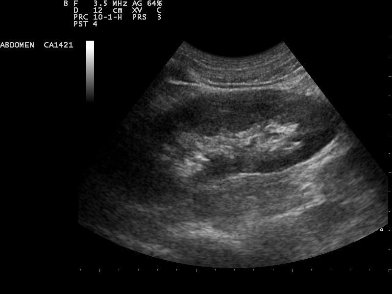
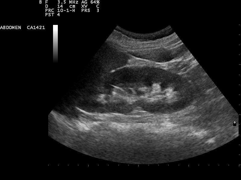
A kidney and bladder ultrasound, or renal ultrasound, uses high frequency sound waves to visualize and assess your kidneys, ureters and urinary bladder. For both men and women, this exam can help detect fluid collection, kidney or urinary tract infection, cysts, tumors, kidney disease, obstructions like kidney stones, and more.
While the severity of bladder and kidney conditions vary, many of them are very common. Its estimated between 40 to 60 percent of women develop a urinary tract infection during their lifetime, and the likelihood of an infection increases as you age. Kidney disease, on the other hand, affects one in 10 Canadians according to the Kidney Foundation of Canada.
Ultrasound imaging, also known as sonography, is often requested when investigating these bladder and kidney concerns, because its very good a looking at the soft tissues of the body, as well as evaluating blood flow and fluid retention.
What Causes Kidney Malfunction
A major culprit of kidney problems is an acidic diet . A brand-new study sheds light on the renal problems that can be caused by a high-acid, meat-rich diet.
The study followed 1,500 people with kidney disease for a period of 14 years. Participants who ate a diet high in meat came very close to experiencing complete kidney failure, while those who ate more fruits and vegetables did not even come close to kidney failure. Researchers estimate that an acidic diet can make it three times more likely for your kidneys to fail.1
Says lead study author Dr. Tanushree Banejee,
Patients with chronic kidney disease may want to pay more attention to diet consumption of acid rich foods to reduce progression to kidney failuredialysis treatmentsmay be avoided by adopting a more healthy diet that is rich in fruits and vegetables.1
What Does The Treatment Involve
You will be positioned on an operating table. A soft, water-filled cushion may be placed on your abdomen or behind your kidney. The body is positioned so that the stone can be targeted precisely with the shock wave. In an older method, the patient is placed in a tub of lukewarm water. About 1-2 thousand shock waves are needed to crush the stones. The complete treatment takes about 45 to 60 minutes.
Sometimes, doctors insert a tube via the bladder and thread it up to the kidney just prior to SWL. These tubes are used when the ureter is blocked, when there is a risk of infection and in patients with intolerable pain or reduced kidney function.
After the procedure, you will usually stay for about an hour then be allowed to return home if all goes well. You will be asked to drink plenty of liquid, strain your urine through a filter to capture the stone pieces for testing, and you may need to take antibiotics and painkillers. Some studies have reported stones may come out better if certain drugs are used after SWL.
Recommended Reading: Is Pineapple Good For Kidney Stones
How Ultrasound Scans Work
A small device called an ultrasound probe is used, which gives off high-frequency sound waves.
You can’t hear these sound waves, but when they bounce off different parts of the body, they create “echoes” that are picked up by the probe and turned into a moving image.
This image is displayed on a monitor while the scan is carried out.
Why Do I Have Pain In My Left Ovary

According to VeryWellhealth.com, ovary pain, which is often felt in the lower abdomen, pelvis, or lower back, are related to ovulation and menstruation. A GYN problem like endometriosis or pelvic inflammatory disease, or even a medical condition affecting your digestive or urinary system can be to blame.
Also Check: Can Kidney Stones Cause Constipation Or Diarrhea
So Now Is The Time To Pamper Your Kidneys
If youre experiencing any of the symptoms mentioned above, you should see your health practitioner and have a check-up, including tests to assess your kidney function.
And you should also make sure youre helping your kidneys stay in top shape by doing a thorough cleanse.
Even if you dont have any of these signs, a systemic cleanse is like a spa day for your kidneys. It helps them get in top shape and avoid damage and disease. This is why OsteoCleanse, The 7 Day Bone Building Accelerator was developed in conjunction with the Osteoporosis Reversal Program.
OsteoCleanse is not just about alkalizing your body, feeling younger and more energized, and removing osteoporosis drugs from your system. It does all of these things in just seven days, but at the heart of OsteoCleanses effectiveness are its kidney-boosting, liver-cleansing effects so youll strengthen and build your bones faster.
Its always a good idea to heed early warning signs and treat your kidneys to a cleanse before damage occurs, and its particularly important to offset the effects of aging on your renal system.
What Does Imaging Mean
Imaging is a general term for techniques used to create pictures. In medicine, imaging produces pictures of bones, organs, and vessels inside the body. Imaging helps health care professionals see the cause of medical problems. Imaging techniques include
- hydronephrosis, or urine blockage, in newborns following suspicious or abnormal imaging during the pregnancy
Don’t Miss: Pineapple For Kidney
What Are The Limitations Of Abdominal Ultrasound Imaging
Ultrasound waves are disrupted by air or gas. Therefore, ultrasound is not an ideal imaging technique for the air-filled bowel or organs obscured by the bowel. Ultrasound is not as useful for imaging air-filled lungs, but it may be used to detect fluid around or within the lungs. Similarly, ultrasound cannot penetrate bone, but may be used for imaging bone fractures or for infection surrounding a bone.
Large patients are more difficult to image by ultrasound because greater amounts of tissue weaken the sound waves as they pass deeper into the body and need to return to the transducer for analysis.
Are There Any Risks Or Side Effects
There are no known risks from the sound waves used in an ultrasound scan. Unlike some other scans, such as CT scans, ultrasound scans don’t involve exposure to radiation.
External and internal ultrasound scans don’t have any side effects and are generally painless, although you may experience some discomfort as the probe is pressed over your skin or inserted into your body.
If you’re having an internal scan and are allergic to latex, it’s important to let the sonographer or doctor carrying out the scan know this so they can use a latex-free probe cover.
Endoscopic ultrasounds can be a bit more uncomfortable and can cause temporary side effects, such as a sore throat or bloating.
There’s also a small risk of more serious complications, such as internal bleeding.
Page last reviewed: 28 July 2021 Next review due: 28 July 2024
Read Also: Constipation And Kidney Stones
Why Wait Until Your Kidneys Are Diseased
While the study was conducted on people with kidney disease, we could safely extrapolate the recommendations to those who want to avoid kidney disease and achieve optimal kidney function now, especially as we age.
In fact, additional research points to the actuality of physiological changes in the kidneys as we age. The research notes that a progressive reduction of the glomerular filtration rate and renal blood flow are observed in conjunction with aging. The reason for these phenomena is a decrease in the plasma flow in the glomerulus, a bundle of capillaries that partially form the renal corpuscle.2
In addition, the aging kidneys experience other structural changes, such as a loss of renal mass, and decreased responsiveness to stimuli that constrict or dilate blood vessels. The study concludes with a notable summation:
age-related changes in cardiovascular hemodynamics, such as reduced cardiac output and systemic hypertension, are likely to play a role in reducing renal perfusion and filtration. Finally, it is hypothesized that increases in cellular oxidative stress that accompany aging result in endothelial cell dysfunction and changes in vasoactive mediators resulting in increased atherosclerosis, hypertension and glomerulosclerosis.2
Are There Different Kinds Of Blockages
Yes. Blockages may occur at the point where the ureter leaves the kidney pelvis, or at the point where the bladder empties into the urethra. These urinary tract abnormalities may be associated with urinary tract infections in children, which can result in kidney injury. However, when detected early and treated appropriately, kidney injury may be avoided in many cases.
Also Check: Cranberry Juice Good For Liver
Left Kidney Ultrasound Transverse View
- Maintaining the longitudinal view, center the kidney on your screen, and then rotate your probe 90 degrees counterclockwise.
- The indicator should be pointing Anteriorly.
- Tilt the probe superiorly and inferiorly to assess the entire left kidney.
- Visualize the same renal structures you did in the Right Kidney.
Like this Post?Sign Up For POCUS 101 Updates!
If You Have Questions

If you have questions about the renal ultrasound, speak with your doctor. You can also talk to the technician before the exam.
Reviewed by: Yamini Durani, MDDate reviewed: March 2012
Note: All information on KidsHealth is for educational purposes only. For specific medical advice, diagnoses, and treatment, consult your doctor.
Don’t Miss: Is Pomegranate Juice Good For Your Kidneys
What Happens During My Renal Ultrasound
- The sonographer may ask you to change into a gown, depending on which organ we are examining.
- Alternatively, the sonographer may arrange your clothing to expose the area of interest so they can apply ultrasound gel.
Important Notes
Since a renal ultrasound focuses on bladder draining, post-void residual, urine flow through the ureters, etc., it is extremely important that you prepare correctly and drink water before your exam. Please visit our exam prep page for specific instructions relating to renal ultrasound.
Orientation
Cost
Duration
A renal ultrasound scan lasts approximately 30 minutes.
What Can I Expect For My Child
Babies with urinary tract abnormalities detected by prenatal ultrasound often do very well. Nevertheless, babies with these conditions need careful evaluation following birth to see if treatment is necessary. Your baby may only need periodic visits to your doctor, or a specialist in the treatment of children with congenital urinary tract abnormalities. Sometimes, a dose of antibiotics at bedtime is prescribed. Occasionally, infants with urinary tract abnormalities may need an operation to correct the problem. Your doctor can provide you with further information regarding congenital urinary tract abnormalities.
If you would like more information, please contact us.
COVID-19 patients can become kidney patients.
You can provide lifesaving support today with a special monthly gift.
Recommended Reading: Palo Azul For Kidney Stones
What Are The Reasons For A Kidney Ultrasound
A kidney ultrasound may be used to assess the size, location, and shapeof the kidneys and related structures, such as the ureters and bladder.Ultrasound can detect cysts, tumors, abscesses, obstructions, fluidcollection, and infection within or around the kidneys.Calculi of the kidneysand ureters may be detected by ultrasound.
A kidney ultrasound may be performed to assist in placement of needlesused tobiopsy the kidneys, to drain fluid from a cyst or abscess, or to place a drainage tube.This procedure may also be used to determine blood flow to the kidneysthrough the renal arteries and veins.
Kidney ultrasound may be used after akidney transplantto evaluate the transplanted kidney.
There may be other reasons for your physician to recommend a kidneyultrasound.
Our Approach to Kidney Ultrasounds
The Johns Hopkins Kidney Program is one of the first in the country and our doctors have pioneered some of the most innovative treatments for patients with renal failure. In addition to offering leading-edge procedures, our program offers shorter wait times and minimally invasive options, which can lead to a faster recovery.
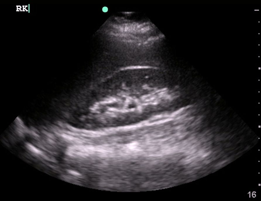
Right Kidney Ultrasound Transverse View
- Maintaining the longitudinal view of the right kidney, center the kidney on your screen, and then rotate your probe 90 degrees counterclockwise.
- The probe indicator should be pointing Posteriorly.
- Here is the ultrasound image transition that you should see as you go from the longitudinal kidney ultrasound view to the transverse view.
Also Check: Celery Juice And Kidney Disease
What To Expect During A Liver Ultrasound
To prepare for your ultrasound, please follow these steps:
- Do not eat any fatty or gassy foods for at least 24 hours before the procedure.
- Avoid eating for eight to 12 hours before your appointment. You may have small sips of water.
- If you need to take regular medication, do so with a small amount of water.
Your abdominal ultrasound will be a simple, pain-free outpatient procedure. During it, the technologist will have you lie down on the table, where theyll apply the ultrasound gel to the area of your abdomen being scanned. The gel helps eliminate air pockets and lets the scanner move more smoothly across your body to pick up clearer images. From there, your technologist will take your ultrasound using a hand-held transducer, the tool that uses sound waves to produce the internal images of your liver.
The scan will feel like a smooth, gentle pressure moving across your abdomen without causing any pain. When the scanning process is over, the team will review your images to check if theyre clear. If a biopsy is required, it might be performed next. The ultrasound process should take about 30 minutes.
After your abdominal ultrasound, your doctor will examine the scanned images, share your results and help you understand both your condition and the next necessary steps. If you have any additional questions about your upcoming liver ultrasound, contact Dr. Mark Fraimans interdisciplinary team today.
This article was medically reviewed by Dr. Mark Fraiman on March 7, 2019.
How Do I Prepare For A Kidney Ultrasound
The preparation for this test will depend on the type of ultrasound procedure your healthcare provider has ordered. Some things you might need to do to get ready for your ultrasound could include:
- Drinking a quart of water before the test to obtain better images.
- Eating a fat-free dinner the night before the test.
- Fasting .
In some cases, you may not need to do anything before your ultrasound. Your healthcare provider will let you know exactly what you need to do before the test. If you have any questions leading up to your ultrasound appointment, call your providers office to learn more about how to prepare.
Also Check: Watermelon Kidney Disease
What Should I Expect
The procedure is short and painless, causing no discomfort to the patient. It is performed in the office. You will not need to undress for the exam. You will be asked to lie down. Your shirt is then pulled up to expose the abdomen and towels are used to protect your clothing from the ultrasound gel. Ultrasound gel is water-based and does not harm the skin or clothing. The physician will then place the cold gel on your abdomen and apply gentle pressure with the ultrasound instrument. The exam should take between 15-30 minutes.
What Does The Equipment Look Like

Ultrasound machines consist of a computer console, video monitor and an attached transducer. The transducer is a small hand-held device that resembles a microphone. Some exams may use different transducers during a single exam. The transducer sends out inaudible, high-frequency sound waves into the body and listens for the returning echoes. The same principles apply to sonar used by boats and submarines.
The technologist applies a small amount of gel to the area under examination and places the transducer there. The gel allows sound waves to travel back and forth between the transducer and the area under examination. The ultrasound image is immediately visible on a video monitor. The computer creates the image based on the loudness , pitch , and time it takes for the ultrasound signal to return to the transducer. It also considers what type of body structure and/or tissue the sound is traveling through.
Also Check: Does Pop Cause Kidney Stones
Can An Ultrasound Diagnose Kidney Disease
May 12, 2016.
Chronic kidney disease diagnosis and staging are based on.
Assays for albumin can be standardized.
or biopsy of the kidney. For example, people with family history of polycystic kidney disease should undergo ultrasound examination to detect.
Kidney cancer is usually diagnosed using a combination of ultrasound, CT, and MRI scans, along with a careful history, physical exam, blood tests, and urine tests. Once a cancer is diagnosed, the tumor needs to be carefully staged in order to determine the most appropriate treatments.
Ultrasound imaging does not use any injected dyes or radiation and is safe for all patients, including pregnant women. It can also detect cysts in the kidneys of a.
Inflammatory disorders of the canine pancreas can.
diagnose acute pancreatitis.8 Laboratory parameters that are important to rule out other organ diseases than pancreatitis and to assess disease.
A kidney ultrasound is an imaging test that allows your healthcare provider to look at your right and left kidney, as well as your bladder. The kidneys are the filtration system of your body. They filter the waste products out of your blood. The waste products then leave your body as urine.
Prediction of kidney function and chronic kidney disease through.
The main clinical application of kidney ultrasound imaging involves excluding.
Furthermore, it can reliably determine whether eGFR is below 60.
Ultrasound findings can be normal in patients with renal disease,