What Are The Signs That Something Is Wrong With Your Kidneys
Signs of Kidney Disease
- Youâre more tired, have less energy or are having trouble concentrating. â¦
- Youâre having trouble sleeping. â¦
- You have dry and itchy skin. â¦
- You feel the need to urinate more often. â¦
- You see blood in your urine. â¦
- Your urine is foamy. â¦
Donât Miss: How To Reverse Kidney Disease Naturally
Anatomical And Surgical Abnormalities
Blockage, or obstruction of the ureter can occur, as a result of narrowing within the ureter, or compression or fibrosis of structures around the ureter. Narrowing can result of ureteric stones, masses associated with cancer, and other lesions such as endometriosistuberculosis and schistosomiasis. Things outside the ureters such as constipation and retroperitoneal fibrosis can also compress them. Some congenital abnormalities can also result in narrowing or the ureters. Congenital disorders of the ureter and urinary tract affect 10% of infants. These include partial or total duplication of the ureter , or the formation of a second irregularly placed ureter or where the junction with the bladder is malformed or a ureterocoele develops . If the ureters have been resited as a result of surgery, for example due to a kidney transplant or due to past surgery for vesicoureteric reflux, that site may also become narrowed.
Pearls And Other Issues
Ureteral injuries are rare, but a high level of suspicion for these injuries is necessary when performing an operation with high risk for ureteral injury because immediate identification of the injury is the most important prognostic factor. If a ureteral injury is suspected, an RPG can be obtained at the time of the procedure. Another method to identify the injury involves direct inspection of the ureters using intravenous dyes such as methylene blue and indigo carmine to evaluate for dye leakage along the course of the ureter. Injury treatment will vary on the location and extent of the injury. Treatment can be as simple as ureteral stent placement and as complicated as using a portion of ileum as ureter replacement.
Recommended Reading: How Do You Diagnose Kidney Stones
How Does The Urinary System Clean My Blood
Your kidneys are an essential part of filtering your blood. Heres how the urinary system works:
Anomalies Of The Ureters

- A blockage may occur in the area where the kidney attaches to the ureter .
- A blockage may occur in the area where the ureter attaches to the bladder .
- The ureters may attach to the bladder in a way that allows urine to flow back up to the kidney .
- A single kidney may drain to the bladder with two ureters instead of one.
Recommended Reading: What Does A Kidney Biopsy Consist Of
What Keeps The Kidneys In Place
Each kidney is held in place by connective tissue, called renal fascia, and is surrounded by a thick layer of adipose tissue, called perirenal fat, which helps to protect it. A tough, fibrous, connective tissue renal capsule closely envelopes each kidney and provides support for the soft tissue that is inside.
What Are The Different Types Of Renal Hilum Trauma
The main renal vessels can be interrupted by thrombosis or avulsion. In both instances, there will be no contrast enhancing the injured kidney in the CAT scan with IV contrast. Hematuria may not be present in these cases. The renal artery injury is often stretched with blunt trauma and an intimal flap may cause obstruction and ischemia. Main renal artery trauma with intimal flap can be successfully managed with arterial endovascular stent placement by interventional radiology.
You May Like: Can You Get Kidney Failure From Drinking Too Much Water
Where Does The Ureter Penetrate The Kidney Quizlet
Where does the ureter penetrate the kidney? The ureter, blood vessels, and nerves penetrate the kidney on its medial surface. The fibrous capsule is a layer of adipose tissue that surrounds the kidney.
Where is the renal vein located in the kidney?
The renal veins lie ventral to the renal arteries. The left is longer than the right and passes in front of the aorta, just inferior to the origin of the superior mesenteric artery. The renal veins enter the vena cava around the level of L1-L2. The left renal vein is usually slightly more cephalad than the right.
Structure The Kidneys Are Bean
Gerotas fascia is a thin, fibrous tissue on the outside of the kidney. Below Gerotas fascia is a layer of fat.
The renal capsule is a layer of fibrous tissue that surrounds the body of the kidney inside the layer of fat.
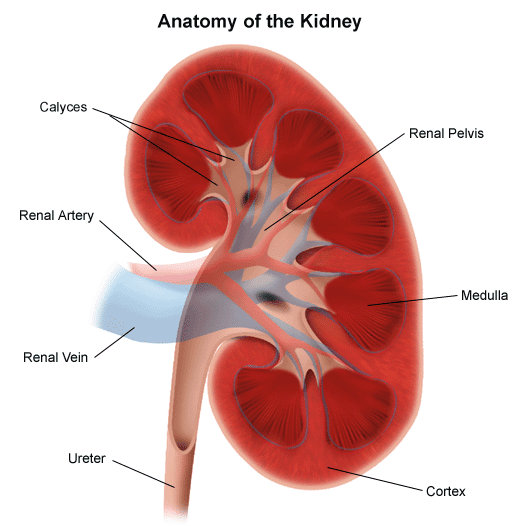
The cortex is the tissue just under the renal capsule.
The medulla is the inner part of the kidney.
The renal pelvis is a hollow area in the centre of each kidney where urine collects.
The renal artery brings blood to the kidney.
The renal vein takes blood back to the body after it has passed through the kidney.
The renal hilum is the area where the renal artery, renal vein and ureter enter the kidney.
The nephrons are the millions of small tubes inside each kidney. Each nephron has 2 parts. Tubules are tiny tubes that collect the waste materials and chemicals from the blood moving through the kidney. The corpuscles contain a clump of tiny blood vessels called glomeruli that filter the blood as it moves through the kidney. The waste products are passed through the tubules to the collecting ducts, which drain into the renal pelvis.
Don’t Miss: Can Kidneys Heal On Their Own
What Is The Kidney Attached To
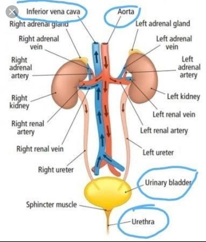
Your kidneys are shaped like beans, and each is about the size of a fist. They are near the middle of your back, one on each side of your spine, just below your rib cage. Each kidney is connected to your bladder by a thin tube called the ureter.
What is the proper path of urine in our body?
Urine is formed in the kidneys by filtering the blood. Urine is then carried through the ureters to the bladder, where it is stored. During urination, urine is passed from the bladder through the urethra to the outside of the body.
What problems can a duplex kidney cause?
Duplex kidney can cause urine to flow back into the kidney instead of the bladder and can also cause urine obstruction.
Where is the kidney in the human body front or back?
The kidneys are a pair of bean-shaped organs on either side of your spine, below your ribs, and behind your belly. Each kidney is about 4 or 5 inches long, about the size of a large fist. The role of the kidneys is to filter your blood.
How Are Most Ureters Damaged
The ureter is the least-often injured genitourinary organ, accounting for less than 1% of all urologic traumas secondary to external violence. Ureteral injury occurs more commonly intraoperatively than from external violent trauma . Blunt trauma and stab wounds rarely result in injury to the ureter. Blunt trauma represents 4.1% and SWs 5.2% of all ureteral traumas. The most common ureteral injuries from external violence are from gunshot wounds .
Read Also: Which Is Not Correct Regarding The Kidneys
Anatomical Features In Equids
Introduction
The following material covers the relevant clinical anatomy of the urinary bladder, including ureters and urethra, in the female and male horse, along with its blood supply, lymphatics, and innervation. Additional information on the equine urinary bladder is also presented in Case 13.10.
Function
The urinary bladder is a hollow muscular organ that collects and stores urine produced by the kidneys. Urine is typically voided from the bladder during urination, a complex physiologic process involving sympathetic, parasympathetic, and voluntary nervous control. Horses typically urinate about every 4 h and produce 515 L of urine per day. The bladder has a capacity of 34 L of urine. The calcium carbonate crystals in horse urine give it a milky yellow appearance. Sometimes, the urine appears reddish-orange in color due to the oxidation of the naturally occurring urocatechin by light.
People often mistake this normal discoloration of the urine for hematuria, especially during the winter months when the urine is seen in the snow.
Urinary bladder
Figure 13.11-3. Lateral view of the female genitourinary tract. Figure inlays are the male and female trigone of the urinary bladder.
What Is The Function Of The Peritubular Capillaries

Peritubular capillaries help your urinary system get rid of waste. They move waste and excess water through your kidneys nephrons . The waste travels into your bladder and leaves your body through urine .
Peritubular capillaries also reabsorb substances your body needs to work properly, such as amino acids, minerals and glucose . They supply blood and oxygen to the cells in a system of tubes in your kidneys as well.
You May Like: How To Get Rid Of Simple Kidney Cysts
What Is Urine Made Of
Urine is made of water, urea, electrolytes, and other waste products. The exact contents of urine vary depending on how much fluid and salt you take in, your environment, and your health. Some medicines and drugs are also excreted in urine and can be found in the urine.
*Electrolytes
As mentioned prior, urine is formed in the nephrons by a three-step process: glomerular filtration, tubular reabsorption, and tubular secretion. The amount of urine varies based on fluid intake and ones environment.
Urine Collection And Emission
From the nephrons the urine enters the final 15 or 20 collecting tubules that open on to each papilla of the renal medulla, projecting into a minor calyx. These open into two or three major calyxes, and these in turn open into the renal pelvis, which connects with the upper expanded portion of the ureter.
Urine is passed down the channel of the renal pelvis and ureter by a succession of peristaltic waves of contraction that begin in the muscle fibres of the minor calyxes, travel out to the major calyxes and then along the ureter every 1015 seconds. Each wave sends urine through the ureteric orifice into the bladder in discontinuous spurts these can be seen through a cystoscope if a dye is injected into the bloodstream. Gravity aids this downward flow, which is faster when one is standing erect. Though the overall picture suggests that there is a pacemaker near the pelviureteric junction, this has never been satisfactorily demonstrated in the tissue. The pressure in the renal pelvis is normally low, but the smooth muscle coat of the ureter is a powerful one and the pressure above an obstructed ureter may rise as high as 50 millimetres of mercury. The ureters are doubly innervated from the splanchnic nerves above and the hypogastric network below.
You May Like: Do Walnuts Cause Kidney Stones
Is The Ureter Attached To The Kidney
The ureter is a tube that carries urine from the kidney to the urinary bladder. There are two ureters, one attached to each kidney. The upper half of the ureter is located in the abdomen and the lower half is located in the pelvic area.
What attaches the kidneys to the Retroperitoneum?
The T12 and L3 vertebrae attach the kidneys to the retroperitoneum.
Where is the urethra and ureter located?
What Are the Ureter and Urethra? The ureter is a small tube, or duct, that connects the bladder and kidneys. Urine passes through the ureter from the kidneys to the bladder. The urethra is the tubular path that connects the bladder to the bodys exterior, allowing urine to exit the body.
What blood vessels receives blood from the renal veins?
The renal veins are veins that drain the kidney. They connect the kidney to the inferior vena cava. They carry the blood filtered by the kidney.
| Renal vein |
|---|
Also Check: How Long Can You Live With Total Kidney Failure
What Do The Kidneys Do
Kidneys have many jobs, from filtering blood and making urine to keeping bones healthy and making a hormone that controls the production of red blood cells. The kidneys also help regulate blood pressure, the level of salts in the blood, and the acid-base balance of the blood. All these jobs make the kidneys essential to keeping the body working as it should.
Also Check: Where Does It Hurt If You Have Kidney Stones
When Should I Call My Doctor If I Think I Might Have A Problem With My Urinary Tract
If youre having trouble or pain when urinating, you should visit your doctor. It may be a sign of an infection or another condition. Call your doctor if you have:
- Blood in your urine.
- Burning sensation, pain or difficulty urinating.
- Pain in your pelvic area, lower back, genital area, or flank .
- Trouble holding your urine or problems with leaking urine.
A note from Cleveland Clinic
Your urinary system plays a critical role in keeping you alive. It filters your blood and removes waste and excess water through urine. Your urinary system includes your kidneys, ureters, bladder and urethra. Conditions like urinary tract infections, sexually transmitted diseases, kidney diseases, and urinary tract obstruction can affect the health of your urinary system. If you have one of these conditions, talk to your healthcare provider about steps you can take to ensure your health.
Last reviewed by a Cleveland Clinic medical professional on 12/05/2019.
References
How Do You Treat A Duplex Kidney
Treatments for duplex kidneysNephrectomy ? kidney removal. Heminephrectomy ? part of the affected kidney and duplicated ureter are removed. Ureteroureterostomy ? in the case of an ectopic ureter, it is split near the bladder and joined to the normal ureter, allowing urine from the upper kidney to drain as normal.
Don’t Miss: What Does 15 Percent Kidney Function Mean
Where Are The Kidneys Located In The Male Body
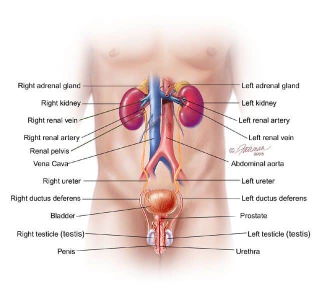
Male urinary system. Male urinary system Your urinary system ? which includes the kidneys, ureters, bladder and urethra ? is responsible for removing waste from your body through urine. Your kidneys, located toward the back in your upper abdomen, produce urine by filtering waste and fluid from your blood.
Duplication Of The Ureter

Ureteral duplication is one of the most common congenital abnormalities of the ureter. Autopsy series have estimated the incidence to be 0.8% or 0.9%.
Ureteral duplication ranges from Y duplication to complete duplication. The Y duplication likely represents divergence of the ureteric bud before meeting the metanephric blastema during development, whereas complete duplication reflects 2 ureteric buds developmentally. Clinically, the incidence of the Y variant is lower than 0.9%, as this duplication is often without consequence. Unilateral ureteral duplication is 6 times more common than bilateral duplication and has no clear sex preponderance or laterality preference. However, a hereditary role in ureteral duplication is evident.
One of the paramount anatomic relationships of duplicated ureters is dictated by the Weigert-Meyer law, which states that the orifice of the lower pole ureter occupies a more cranial and lateral position. In general, the upper pole ureter drains less renal parenchyma. The lower pole ureter is more susceptible to dilatation from vesicoureteral reflux than single ureters or upper pole ureters. The upper pole ureters are often subject to anomalies of termination such as ureterocele or ectopic orifice.
You May Like: Where Is Back Pain Located For Kidney Infection
What Are The Kidneys And Urinary Tract
The urinary tract is one of the systems that our bodies use to get rid of waste products. The kidneys are the part of the urinary tract that makes urine . Urine has salts, toxins, and water that need to be filtered out of the blood. After the kidneys make urine, it leaves the body using the rest of the urinary tract as a pathway.
What Are The Treatment Options For Urinary Leakage After Renal Trauma
Urinary extravasation usually does not require surgical intervention. Initially these patients should be managed with bladder drainage to decompress the upper urinary tract. In cases of penetrating injury and gunshot trauma, the ureter can suffer a blast effect that can result in delayed urine extravasation in 48â72 hours, with urinoma formation. In these cases, ureteral stents with or without percutaneous drainage of the urine collection can effectively manage this injury. Surgical repair should be reserved for patients who failed minimally invasive treatments.
Donât Miss: Do Kidney Stones Hurt When You Move
Don’t Miss: Which Side Is Your Kidney On
What Is The Junction Between The Kidney And The Bladder Called
Structure. The junction between the kidney pelvis and the ureters is known as the uretero-pelvic junction or ureteral pelvic junction, and the junction between the ureter and the bladder is known as the uretero-vesical junction . At the entrance to the bladder, the ureters are surrounded by so-called uretero-vesical valves
What is the tube that carries urine to the bladder?
The ureters are tubes that carry urine and connect the kidneys to the bladder. 1. Human urinary system: 2. Kidney, 3. Renal pelvis, 4. Ureter, 5.
Where are the kidneys located in the male body?
Male urinary system. Male Urinary System Your urinary system which includes the kidneys, ureters, bladder, and urethra is responsible for removing waste from your body through urine. Your kidneys, located towards the back in your upper abdomen, produce urine by filtering waste and fluids from your blood.
Where Does The Ureters Lead To
The urine travels from the kidneys to the bladder in two thin tubes called ureters. The ureters are about 8 to 10 inches long. Muscles in the ureter walls tighten and relax to force urine down and away from the kidneys. Small amounts of urine flow from the ureters into the bladder about every 10 to 15 seconds.
Also Check: How To Detect Kidney Infection