Major Calyx Disease Diagnosis Of Caliectasis
The diagnostic procedures for Caliectasis include:
- Physical exam A thorough physical examination is performed by the doctor to understand if there is any tenderness or swelling around the kidney area.
- Urinalysis It is a test done by taking a urine sample.
- Ultrasound Doctors perform an abdominal ultrasound test to diagnose any extra fluids collected in the kidneys or if there are any foreign objects present there.
- Urography The procedure uses contrast dye and a CT scan to get a clear view of your kidneys.
- Cystoscopy A urethra is inserted inside your body with a camera at its one end to take pictures of the inside of the bladder and your kidneys.
Capillary Network Within The Nephron
The capillary network that originates from the renal arteries supplies the nephron with blood that needs to be filtered. The branch that enters the glomerulus is called the afferent arteriole. The branch that exits the glomerulus is called the efferent arteriole. Within the glomerulus, the network of capillaries is called the glomerular capillary bed. Once the efferent arteriole exits the glomerulus, it forms the peritubular capillary network, which surrounds and interacts with parts of the renal tubule. In cortical nephrons, the peritubular capillary network surrounds the PCT and DCT. In juxtamedullary nephrons, the peritubular capillary network forms a network around the loop of Henle and is called the vasa recta.
Major Calyx Minor Calyx Renal Capsule Anatomy In The Kidney
Major calyx, minor calyx, renal capsule anatomy in the kidney
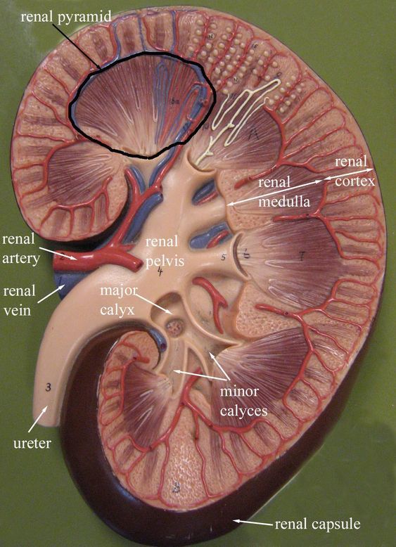
In this image, you will find Medulla, Cortex, Renal pyramid, Connection for minor calyx, Minor calyx, Major calyx, Renal lobe, Renal columns, Renal capsule, Renal papilla, Ureter, Hilus, Renal pelsiv, Adipsope tissue in renal sinus, Renal sinus it.
We are pleased to provide you with the picture named Major calyx, minor calyx, renal capsule anatomy in the kidney. We hope this picture Major calyx, minor calyx, renal capsule anatomy in the kidney can help you study and research. for more anatomy content please follow us and visit our website: www.anatomynote.com.
Anatomynote.com found Major calyx, minor calyx, renal capsule anatomy in the kidney from plenty of anatomical pictures on the internet. We think this is the most useful anatomy picture that you need. You can click the image to magnify if you cannot see clearly.
This image added by admin. Thank you for visit anatomynote.com. We hope you can get the exact information you are looking for. Please do not forget to share this page and follow our social media to help further develop our website. If you have any question please do not hesitate to contact us.
If you think this picture helpful, please don’t forget to rate us below the picture!
One of our purpose to collect these pictures is we hope these pictures will not be lost when the relevant web page is deleted.
Major calyx, minor calyx, renal capsule anatomy in the kidney
Also Check: Is Grape Juice Good For Kidneys
Kidney Disease And Disorders
Kidney diseases and kidney problems are usually treated by a nephrologist. Kidney stones are sometimes treated by a urologist. Here is a list of some of the more common kidney problems:
- Glomerulonephritis inflammation of the glomeruli
- Hydronephrosis excessive fluid within the kidney caused by blocked urine flow
- Pyelonephritis infection of the kidney
- Kidney Stones usually form in the kidneys, but can form anywhere in the urinary tract
- Kidney Cancer
- Nephrosis a process that can lead to kidney failure
- Polycystic Kidney Disease a disorder of the kidneys that result in multiple fluid filled cysts within the kidneys tissues
- Renal Hypertension if the kidneys for some reason do not get enough blood, they set off a series of events leading to high blood pressure
- Renal Infarction similar to a heart attack, but in the kidney, caused by blockage of kidney vessels
- Renal Vein clot clot in the vein that carries blood from the kidney, can be fatal
What Is Urine Made Of

Urine is made of water, urea, electrolytes, and other waste products. The exact contents of urine vary depending on how much fluid and salt you take in, your environment, and your health. Some medicines and drugs are also excreted in urine and can be found in the urine.
- 94% water
- .1% uric acid
*Electrolytes
As mentioned prior, urine is formed in the nephrons by a three-step process: glomerular filtration, tubular re-absorption, and tubular secretion. The amount of urine varies based on fluid intake and ones environment.
Read Also: Va Rating For Stage 3 Kidney Disease
What Problems Can A Pelvic Kidney Cause
However, one-third of all children born with a pelvic kidney have other complications either with their cardiovascular system, the central nervous system or their urinary system. Symptoms directly associated with the horseshoe kidney can include urinary tract infection, kidney stones or hydronephrosis.
Clinical Relevance: Variation In Arterial Supply To The Kidney
The kidneys present a great variety in arterial supply these variations may be explained by the ascending course of the kidney in the retroperitoneal space, from the original embryological site of formation to the final destination . During this course, the kidneys are supplied by consecutive branches of the iliac vessels and the aorta.
Usually the lower branches become atrophic and vanish while new, higher ones supply the kidney during its ascent. Accessory arteries are common . An accessory artery is any supernumerary artery that reaches the kidney. If a supernumerary artery does not enter the kidney through the hilum, it is called aberrant.
Recommended Reading: What Laxative Is Safe For Kidneys
What Is The Function Of The Renal Papilla
renal papillarenalkidney
. Subsequently, one may also ask, what is the function of the renal pelvis?
The renal pelvis functions as a funnel for urine flowing to the ureter. The renal pelvis is the location of several kinds of kidney cancer and is affected by infection in pyelonephritis.
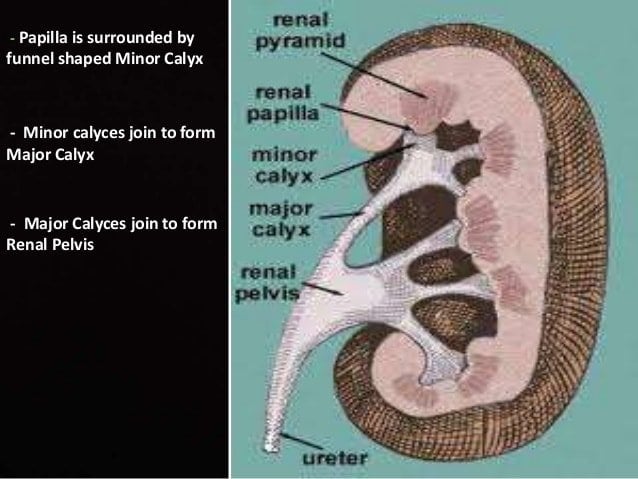
Subsequently, question is, what is the function of the minor calyx? A minor calyx surrounds the renal papillae of each pyramid and collects urine from that pyramid. Several minor calyces converge to form a major calyx. From the major calyces, the urine flows into the renal pelvis and from there, it flows into the ureter.
Besides, what is a renal papilla quizlet?
Projection with tiny openings into a minor calyx. Renal papilla. Superior funnel-shaped end of ureter inside renal sinus. Renal pelvis. relative positions with respect to the architecture of the nephron.
What is renal column?
The renal column is a medullary extension of the renal cortex in between the renal pyramids. It allows the cortex to be better anchored. Each column consists of lines of blood vessels and urinary tubes and a fibrous material.
What Is The Medical Term For Renal Pelvis
Renal pelvis, enlarged upper end of the ureter, the tube through which urine flows from the kidney to the urinary bladder. The large end of the pelvis has roughly cuplike extensions, called calyces, within the kidneythese are cavities in which urine collects before it flows on into the urinary bladder.
You May Like: Is Cranberry Juice Good For Your Liver And Kidneys
What Are The Renal Calyces
Renal calyces are parts of the kidney that collect urine before it passes further into the urinary tract. The calyces are part of the renal pelvis, a convex system of sinuses that connect the innermost part of the kidney to the ureters and, from there, to the bladder. There are two types of renal calyx: the minor calyx and the major calyx. In the human body, there can be two to three major calyces and eight to fourteen minor calyces. Basically, the renal calyces are sectional, hollow cavities that act as reservoirs and help the kidneys perform their duty.
The function of the kidney is to filter out waste and excess water from about 200 quarts of blood every day. Most of this processing takes place in the renal cortex and the renal medulla, the “meaty” parts of the kidney. Once this filtration process is complete, the waste is expunged into the renal calyces and collected. The calyces, branching vessels connected to the kidney interior, empty into the renal pelvis, forming the initial portion of the lower urinary tract.
How Can I Improve My Kidney Function Naturally
Step 5: Stay Healthy
Don’t Miss: Grapes Kidney Stones
Normal Urine Transport And Micturition
The micturition cycle is best thought of as two distinct phases: urine storage/bladder filling and voiding/bladder emptying . The viscoelastic properties of the bladder allow for increases in bladder volume with little change in detrusor or intravesical pressures. Additionally, during bladder filling, spinal sympathetic reflexes are activated that, through modulation of parasympathetic-ganglionic transmission, inhibit bladder contractions and increase bladder-outlet resistance via smooth-muscle activation . Bladder-outlet resistance also increases during filling secondary to increased external urethral-sphincter activity via a spinalsomatic reflex . As the bladder reaches its capacity, afferent activity from tension, volume, and nociceptive receptors are conveyed via A and C fibers through the pelvic and pudenal nerves to the sacral spinal cord . Afferent signals ascend in the spinal cord to the pontine micturition center in the rostral brainstem. Here signals are processed under the strong influence of the cerebral cortex and other areas of the brain. If voiding is deemed appropriate, the voiding/bladder-emptying reflex is initiated. The pattern of efferent activity that follows is completely reversed, producing sacral parasympathetic outflow and inhibition of sympathetic and somatic pathways. First the external urethral-sphincter relaxes and shortly thereafter a coordinated contraction of the bladder causes the expulsion of urine .
What Is Work Of Calyx In Kidney

Answer:
The minor calyces surround the apex of the renal pyramids.
Urine formed in the kidney passes through a renal papilla at the apex into the minor calyx two or three minor calyces converge to form a major calyx, through which urine passes before continuing through the renal pelvis into the ureter.
Peristalsis of the smooth muscle originating in pace-maker cells originating in the walls of the calyces propels urine through the renal pelvis and ureters to the bladder. The initiation is caused by the increase in volume that stretches the walls of the calyces. This causes them to fire impulses which stimulate rhythmical contraction and relaxation, called peristalsis. Parasympathetic innervation enhances the peristalsis while sympathetic innervation inhibits it.The renal calyces are chambers of the kidney through which urine passes. The minor calyces surround the apex of the renal pyramids. Urine formed in the kidney passes through a renal papilla at the apex into the minor calyx two or three minor calyces converge to form a major calyx, through which urine passes before continuing through the renal pelvis into the ureter.
Read Also: What’s A Kidney
Structure Of Human Kidney
- In renal system: Internal configuration
of the cavity called the major calyxes. The major calyxes are divided in turn into four to 12 smaller cuplike cavities, the minor calyxes, into which the renal papillae project. The renal pelvis serves as the initial reservoir for urine, which flows into the sinus through the urinary collecting tubules,
Major Calyx Disease Treatment For Caliectasis
The treatment options chosen by the doctor depends upon the underlying of caliectasis. The several options include:
- Surgery for removal or kidney stones or tumors
- Catheters or nephrostomy tubes to drain urine
- Antibiotics for infections
The condition of Caliectasis is triggered by any underlying problems as traced in the kidneys. Although in most of the cases Caliectasis is often undetected it is often diagnosed when conducting other tests for related kidney issues. The problem, once detected, should be treated right away, otherwise, it may lead to permanent kidney damage.
Recommended Reading: Does Pellegrino Cause Kidney Stones
What Does 10 Percent Kidney Function Mean
It means your kidneys no longer function well enough to meet the needs of daily life. End-stage kidney disease is also called end-stage renal disease . The kidneys of people with ESRD function below 10 percent of their normal ability, which may mean theyre barely functioning or not functioning at all.
Normal Anatomy And Physiology Of The Urinary Tract
The mammalian urinary tract is a contiguous hollow-organ system whose primary function is to collect, transport, store, and expel urine periodically and in a highly coordinated fashion . In so doing, the urinary tract ensures the elimination of metabolic products and toxic wastes generated in the kidneys. The process of constant urine flow in the upper urinary tract and intermittent elimination from the lower urinary tract also plays a crucially important part in cleansing the urinary tract, ridding it of microbes that might have already gained access . When not eliminating urine, the urinary tract acts effectively as a closed system, inaccessible to the microbes. Comprised, from proximal to distal, of renal papillae, renal pelvis, ureters, bladder, and urethra, each component of the urinary tract has distinct anatomic features and performs critical functions.
You May Like: What Std Messes With Your Kidneys
What Happens To The Pleura At The Hilum
The visceral-parietal reflection surrounding the root of the lung extends downwards from the hilum to near the base of the lower lobe in a sleeve-like fold called the pulmonary ligament. At the lower edge of each lung, the pleural layers come into contact with each other, and terminate in a free curved edge.
Microscopic Anatomy And Physiology Of The Urinary Tract
Uroplakins appear to play a major role during the pathogenesis of urinary tract infections.
UPIa presents a high level of terminally exposed, unmodified mannose residues and has been identified as the sole urothelial receptor to interact with the FimH lectin of the type 1-fimbriated uropathogenic E. coli . In addition to the bladder, UPIa has been found on the mucosal surfaces of the ureters, renal pelvis, and major and minor calyces . It has been proposed that interaction of the FimH adhesin of type 1-fimbriated UPEC with UPIa at these locations help bacteria resist the flow of urine and, coupled with bacterial flagella formation, may facilitate the ascent of bacteria from the bladder into the upper urinary tract .
Although both FimH and flagella are known to exhibit phase variation , the temporal expression of these virulence factors in relation to the ascent of UPEC along the urinary tract has not been established. UPEC that cause pyelonephritis typically express P fimbriae in addition to type-1 fimbriae . Once bacteria reach the kidney, the P fimbriae interact with glycolipids in the renal tubular cells removing the need for type-1 fimbriae/UPIa interaction. UPIIIa has also been shown to be important in UTI pathogenesis. Thumbikat et al. demonstrated that the phosphorylation of UPIIIas cytoplasmic tail is a critical step in urothelial signaling associated with bacterial invasion and host-cell apoptosis .
You May Like: Can You Have 4 Kidneys
How Many Major Calyxs Are In The Kidney
4.8/5kidneycalycescalycesrenal
Likewise, how many major Calyces are found in each kidney?
Urine formed in the kidney passes through a renal papilla at the apex into the minor calyx two or three minor calyces converge to form a major calyx, through which urine passes before continuing through the renal pelvis into the ureter.
Also, what is the anatomy of the kidney? Gross AnatomyThe urinary system of the human body consists of two kidneys, two ureters, the bladder and a single urethra. The kidneys are located on the posterior wall of the abdomen at waist level. Each kidney is roughly 10 cm long and 5 cm wide, and is encased in a fibrous outer capsule called the renal capsule.
Secondly, what is the function of the Calyces in the kidney?
function. has roughly cuplike extensions, called calyces, within the kidneythese are cavities in which urine collects before it flows on into the urinary bladder.
Is Extrarenal pelvis dangerous?
An extrarenal pelvis is a normal anatomical variant that is predominantly outside the renal sinus and is larger and more distensible than an intrarenal pelvis that is surrounded by sinus fat. While extrarenal pelvis is asymptomatic in most cases, complications such as infection and stone formation have been reported 2.
What Is The Main Function Of The Calyx

The main function of the calyx is to protect the flower in its bud condition. The calyxes are usually green in colour but sometimes it may be coloured as petalloid, then it can attract insect and helps in pollination. In certain cases calyx develop spur and can store nectar or sometime it helps in seed dispersal too.
You May Like: Can Aleve Damage Your Kidneys
Structure And Function Of The Kidneys
A healthy human being has two kidneys. These are bean-shaped organs located in the rear abdominal cavity, left and right of the spine, slightly beneath the diaphragm under the rib cage, with the left kidney sitting behind the spleen and the right one behind the liver.
The concave border of each kidney has a recessed area that is called the renal hilum. This is where the renal artery enters to supply the kidney with blood carrying oxygene and nutrients, but also waste products, and where the renal vein leaves, carrying the blood that was filtered by the kidney. The hilum is also the entry for lymph vessels, nerves and the ureter. The ureter is a muscular tube that transports the urine from the kidney to the urinary bladder, from where it is released from the body.
Each kidney is encapsulated by a solid cover of connective tissue and embedded in further layers of fat and connective tissue, which protect the organ from trauma and anchor it to the rear abdominal wall
The inner organ can be divided into three major parts: the outer renal cortex, the inner renal medulla and the renal pelvis in the area of the hilum.
Good to know: major function of the kidneys is to filter the blood from toxins and metabolic end products as well as to remove and retain water and electrolytes by making urine, which is released via the ureter and the urinary bladder.
Kidney tissue with nephrons
Overview of the kidneys’ chores