How Much Do My Kidneys Weigh
The weight of your kidneys varies. Variances may include your height, weight, age, body mass index and location.
For men and people assigned male at birth, your right kidney may range from 1/5 to about 1/2 lbs. . Your left kidney may range from a little less than 1/5 to a little more than 1/2 lbs. . Your kidneys may weigh between the weight of one tennis ball and four tennis balls.
For women and people assigned female at birth, your right kidney may range from a little more than 1/10 to 3/5 lbs. . Your left kidney may range from 3/20 to a little less than 3/5 lbs. . Your kidneys may weigh between the weight of one tennis ball or five tennis balls.
How The Kidneys Work
Blood is filtered at high pressure to remove glucose, water, salts and urea.
All the glucose, and some water and salts, are reabsorbed back into the blood. Note that urea is not reabsorbed.
Dr Alice Roberts dissects a pigs kidney and explains the structure and function of the kidney and urinary system
Surgical Indications For Upjhn Based On The Pyeloplasty Prediction Score
A recent study has suggested a pyeloplasty prediction score using three ultrasound parameters to determine who need surgery and who do not in infants with UPJ-like hydronephrosis . They recommend a combination of SFU grade , transverse AP diameter , and the absolute percentage difference of ipsilateral and contralateral renal lengths at baseline to predict a criterion for surgical need. This study suggests that any infant with UPJO-like hydronephrosis with a PPS of 8 or higher is 8 times more likely to undergo pyeloplasty . Unfortunately, none of these parameters is ideal to use due to many disadvantages and/or limitations as described in this review in details. We think that when we put problematic parameters together, it is difficult to get a correct beneficial result from them. Moreover, the laterality , contralateral or bilateral hydronephrosis, ipsilateral atrophy, or contralateral hypertrophy significantly changes the results of the pyeloplasty prediction score . The absolute percentage would be low when there is a contralateral compensatory growth or an atrophy in ipsilateral kidney which will miss the severity of hydronephrosis. In addition, how would it be an objective criterion in bilateral cases? Any of these parameters can change the percentage from 5 to 20%, which means the score may change from 0 to 4. We should use objective and reproducible criteria that are not affected by many parameters and are applicable for all patients.
You May Like: Wine For Kidney Stones
Is The Number Of People With Kidney Cancer Increasing
The incidence of kidney cancer has been increasing nationally over the past thirty years. This is also true in New York State. Despite the increase in incidence, mortality from kidney cancer has decreased slightly over the last 20 years. Much of the increased incidence is in cancers diagnosed at an early stage.
Scientists are trying to determine the reasons for the increase in kidney cancer. Some of the increase may be because health care providers are better able to find the disease through ultrasounds and other medical tests, which can find these cancers at an earlier stage. Some of the increase may also be due to the increasing prevalence of obesity.
How Are Renal Cysts Diagnosed And Evaluated

Since they rarely cause symptoms, renal cysts are most often found during imaging tests performed for other reasons. In such cases without any symptoms, simple renal cysts are usually left alone and do not need any further tests. However, some renal cysts look more complex than the usual simple renal cyst. These complex renal cysts can have a thicker wall, or solid material inside instead of just fluid. Once complex renal cysts are discovered, additional imaging tests may be performed to monitor them and distinguish benign cysts from cancer.
Some types of imaging tests your doctor might order include:
Abdominal Ultrasound and Pelvic Ultrasound: These exams are performed to take pictures of the kidneys and confirm the presence of fluid inside the renal cysts. Your doctor may use ultrasound imaging to monitor renal cysts for any changes over time.
For more information about ultrasound performed on children, visit the Pediatric Abdominal Ultrasound page.
Abdominal and Pelvic CT: Often used as a complement to ultrasound in the study of complex renal cysts, this procedure can help distinguish benign cysts from tumors in the kidneys. A CT scan may include an injection of contrast material. See the Radiation Dose page for more information about CT procedures.
For more information about CT performed on children, visit the Pediatric CT page.
Recommended Reading: Flomax For Kidney Stones In Woman
An Extrarenal Pelvis Is A Normal Variation From The Usual Anatomy And Does Not Necessarily Indicate A State Of Disease
Written by Mansi Kohli | Updated : March 5, 2018 5:48 PM IST
The renal pelvis is a chamber where all the urine-forming ducts meet and further routes urine to the urinary bladder. Any portion of the renal pelvis located outside the kidney is considered as the extrarenal pelvis. Dr Jatin Kothari, Consultant Nephrologist at Hinduja Healthcare Surgical at Khar will help us in understanding the complete concept of the extrarenal pelvis in detail. Also read signs that your kidney is in danger.
What is extrarenal pelvis?
Also known as renal pelvis, it protrudes from the bean-shaped indentation in the middle of the kidney. An extrarenal pelvis is a normal variation from the usual anatomy and does not necessarily indicate a state of disease. Usually, renal pelvis does not protrude in such a manner and it appears as if there is a blockage in the renal pelvis that is preventing urine from emptying.
What Is Urine Made Of
Urine is made of water, urea, electrolytes, and other waste products. The exact contents of urine vary depending on how much fluid and salt you take in, your environment, and your health. Some medicines and drugs are also excreted in urine and can be found in the urine.
- 94% water
- .1% uric acid
*Electrolytes
As mentioned prior, urine is formed in the nephrons by a three-step process: glomerular filtration, tubular re-absorption, and tubular secretion. The amount of urine varies based on fluid intake and ones environment.
Read Also: Is Pomegranate Juice Good For Kidney Stones
Renal Function Following Nephrectomy
Urologic management should focus on optimizing renal function and avoiding CKD whenever possible.
Protection of renal function is a primary concern of physicians who manage surgical or medical diseases of the kidney, such as renal tumors, urinary calculi, renal vascular disease, or ureteropelvic junction obstruction.
Although these disease entities are still managed with nephrectomy in certain situations, approaches that better preserve renal function, including partial nephrectomy, endoscopic stone surgery, angiographic management of renovascular disease, and pyeloplasty are generally preferred.
Many of these patients have pre-existing CKD or are at risk of developing CKD because they also have hypertension, diabetes, systemic atherosclerotic disease, or other comorbid conditions. Therefore preservation of renal function impacts the management of these conditions as well.
Until recently, the main focus of many urologic interventions has been to prevent or delay the need for renal replacement therapy because this end point is associated with increased mortality, as well as a substantial decline in quality of life .
Data also indicate that CKD is independently associated with morbid cardiac events and all-cause mortality in a dose-dependent fashion, even after controlling for a variety of potentially confounding factors such as hypertension and diabetes.
In these studies, approximately 25%-30% of patients with normal SCr levels have at least moderate CKD .
Preparation For A Renal Biopsy
Typically, you dont need to do much to prepare for a renal biopsy.
Be sure to tell your doctor about any prescription drugs, over-the-counter medications, and herbal supplements youre taking. You should discuss with them whether you should stop taking them before and during the test, or if you should change the dosage.
Your doctor may provide special instructions if youre taking medications that could affect the results of the renal biopsy. These medications include:
- anticoagulants
- nonsteroidal anti-inflammatory drugs, including aspirin or ibuprofen
- any medications that affect blood clotting
- herbal or dietary supplements
Tell your doctor if youre pregnant or think you might be pregnant. Also, before your renal biopsy, youll have a blood test and provide a urine sample. This ensures that you dont have any preexisting infections.
You need to fast from food and drink for at least eight hours prior to your kidney biopsy.
If youre given a sedative to take at home before the biopsy, you wont be able to drive yourself to the procedure and need to arrange for transportation.
Recommended Reading: Stds That Affect Kidneys
Diagnosis Of Renal Pelvis And Ureter Cancer
-
Computed tomography or ultrasonography
-
Ureteroscopy
The cancer is usually detected by using computed tomography Computed tomography There are a variety of tests that can be used in the evaluation of a suspected kidney or urinary tract disorder. X-rays are usually not helpful in evaluating… read more or ultrasonography Ultrasonography There are a variety of tests that can be used in the evaluation of a suspected kidney or urinary tract disorder. X-rays are usually not helpful in evaluating… read more . CT and often ultrasonography can help doctors distinguish other noncancerous kidney and ureteral problems such as stones or blood clots. Microscopic examination of a urine sample may reveal cancer cells. A flexible viewing tube with a camera at its tipa ureteroscopethreaded up through the bladder may be used to view cancers, obtain tissue samples for confirmation of the diagnosis, and occasionally even treat small cancers. This is typically done under general anesthesia. To determine how extensive cancers are and how far they have spread, CT scans of the abdomen and pelvis and chest x-ray or CT of the chest are done.
Hormones Of The Adrenal Glands
The role of the adrenal glands in your body is to release certain hormones directly into the bloodstream. Many of these hormones have to do with how the body responds to stress, and some are vital to existence. Both parts of the adrenal glands the adrenal cortex and the adrenal medulla perform distinct and separate functions.
Each zone of the adrenal cortex secretes a specific hormone. The key hormones produced by the adrenal cortex include:
You May Like: Can Mio Cause Kidney Stones
Loss Of Renal Medullary Hypertonicity
- a.
-
The normal hypertonicity of the renal medulla is crucial for elaboration of highly concentrated urine. When medullary hypertonicity is decreased, the osmotic gradient necessary for movement of water from the collecting ducts into the interstitium and back into the bloodstream is disrupted, causing inappropriately dilute urine.
- b.
-
Although the major cause of impaired concentrating ability in chronic renal disease is the necessity to excrete the daily solute load with a decreased number of functional nephrons, several renal diseases are associated with structural abnormalities that also can contribute to impaired concentrating ability.
- c.
-
Renal medullary washout of solute.
-
Long-standing PU/PD of any cause can result in loss of medullary solutes necessary for normal urinary concentrating ability.
-
Structural lesions need not be present for impaired concentrating ability to occur.
-
Increased medullary blood flow associated with long-standing PU/PD can accelerate removal of solutes by the systemic circulation.
-
Aldosterone deficiency in hypoadrenocorticism impairs NaCl reabsorption in the collecting ducts and contributes to medullary washout of solute. This effect explains why dogs with hypoadrenocorticism often have impaired urinary concentrating ability at presentation despite having structurally normal kidneys.
- 1.
Richard W. Nelson, in, 2015
What You Need To Know

- Adrenal glands, also known as suprarenal glands, are small, triangular-shaped glands located on top of both kidneys.
- Adrenal glands produce hormones that help regulate your metabolism, immune system, blood pressure, response to stress and other essential functions.
- Adrenal glands are composed of two parts the cortex and the medulla which are each responsible for producing different hormones.
- When adrenal glands dont produce enough hormones, this can lead to adrenal insufficiency .
- Adrenal glands may develop nodules that can be benign or malignant, which can potentially produce excessive amounts of certain hormones leading to various health issues.
Read Also: Is Watermelon Bad For Your Kidneys
What Causes Kidney Damage
Your kidneys perform several important functions within your body. Many different disorders can affect them. Common conditions that impact your kidneys include:
- Chronic kidney disease: Chronic kidney disease may lessen your kidney function. Diabetes or high blood pressure usually causes CKD.
- Kidney cancer: Renal cell carcinoma is the most common type of kidney cancer.
- Kidney failure : Kidney failure may be acute or chronic . End-stage renal disease is a complete loss of kidney function. It requires dialysis .
- Kidney infection : A kidney infection can occur if bacteria enter your kidneys by traveling up your ureters. These infections cause sudden symptoms. Healthcare providers treat them with antibiotics.
- Kidney stones: Kidney stones cause crystals to form in your urine and may block urine flow. Sometimes these stones pass on their own. In other cases, healthcare providers can offer treatment to break them up or remove them.
- Kidney cysts: Fluid-filled sacs called kidney cysts grow on your kidneys. These cysts can cause kidney damage. Healthcare providers can remove them.
- Polycystic kidney disease: Polycystic kidney disease causes cysts to form on your kidneys. PKD is a genetic condition. It may lead to high blood pressure and kidney failure. People with PKD need regular medical monitoring.
Countless other disorders can affect your kidneys. Some of these conditions include:
Kidney Pelvis And Papilla
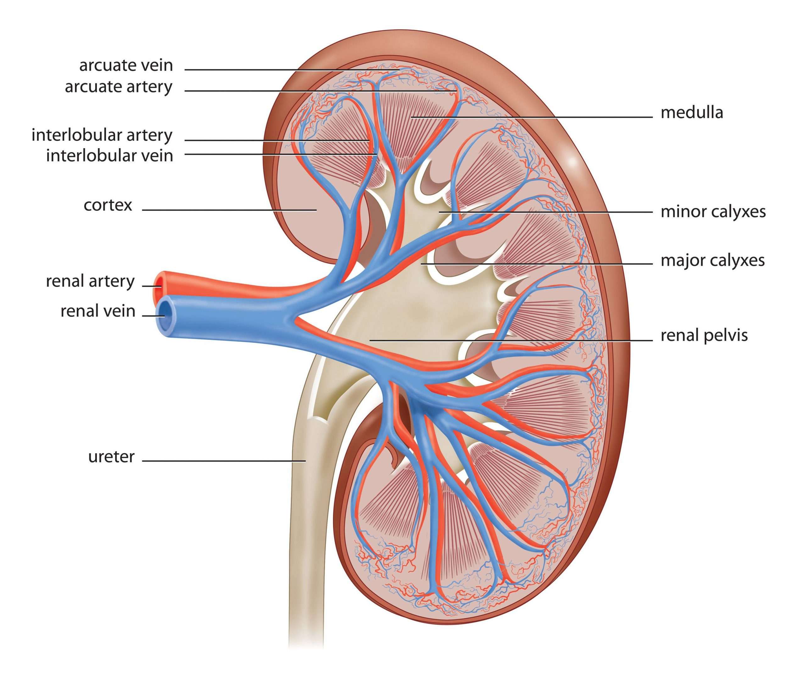
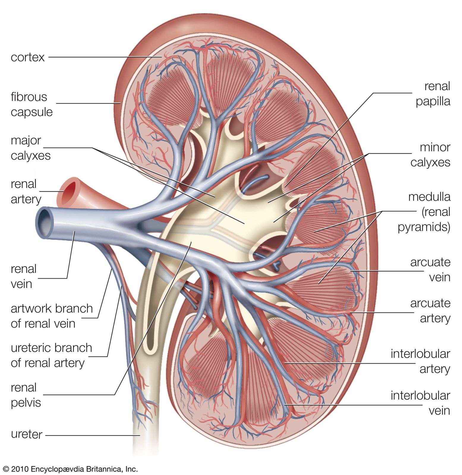
The kidney pelvis acts like a funnel, collecting the urine produced in the kidney and leading to a central stem, the ureter. The epithelial lining of the kidney pelvis is an urothelium, beginning as a single cell layer at the fornices and expanding to three to six cell layers when it reaches the ureter. The lining epithelium of the kidney papilla is a flattened to cuboidal epithelium which does not have the differentiation characteristics of the urothelium. As a consequence, proliferative changes of the lining papilla, such as commonly occur in chronic progressive nephropathy , should not be referred to as kidney pelvis or urothelial hyperplasia, but rather should be referred to as hyperplasia of the lining epithelium of the papilla. Mouse and rat kidneys each have a single papilla projecting into the lumen of the pelvis. Larger mammals, including humans, have multiple papillae.
Samuel M. Cohen, … Shoji Fukushima, in, 2002
Recommended Reading: Watermelon Renal Diet
Kidney And Urinary System Parts And Their Functions
-
Two kidneys. This pair of purplish-brown organs is located below the ribs toward the middle of the back. Their function is to:
-
Remove waste products and drugs from the body
-
Balance the body’s fluids
-
Release hormones to regulate blood pressure
-
Control production of red blood cells
The kidneys remove urea from the blood through tiny filtering units called nephrons. Each nephron consists of a ball formed of small blood capillaries, called a glomerulus, and a small tube called a renal tubule. Urea, together with water and other waste substances, forms the urine as it passes through the nephrons and down the renal tubules of the kidney.
Two sphincter muscles. These circular muscles help keep urine from leaking by closing tightly like a rubber band around the opening of the bladder.
Nerves in the bladder. The nerves alert a person when it is time to urinate, or empty the bladder.
Urethra. This tube allows urine to pass outside the body. The brain signals the bladder muscles to tighten, which squeezes urine out of the bladder. At the same time, the brain signals the sphincter muscles to relax to let urine exit the bladder through the urethra. When all the signals occur in the correct order, normal urination occurs.
Can You Live Without A Kidney
You can live with just one kidney. Healthcare providers may remove one of your kidneys in a radical nephrectomy.
Someone may have only one kidney if they:
- Had a kidney removed due to cancer or injury.
- Made a kidney donation to someone else for a kidney transplant.
- Were born with only one kidney .
- Were born with two kidneys but only one kidney works .
Don’t Miss: Does Chocolate Cause Kidney Stones
Is Drinking A Lot Of Water Good For My Kidneys
Drinking an appropriate amount of water is good for your kidneys. Water helps your kidneys get rid of toxins and wastes through your pee. It also helps keep your blood vessels healthy, making it easier for blood to deliver necessary nutrients to your kidneys.
Its also a good idea to drink an appropriate amount of water to help prevent kidney stones and urinary tract infections . Kidney stones are less likely to form when you have enough water in your kidneys. Youre less likely to get a UTI when you drink a lot of water because youll pee more. Peeing helps flush out the bacteria that cause UTIs.
In general, the color of your pee can reveal if youre drinking enough water. Your pee should be light yellow or clear if youre drinking enough water. If youre dehydrated, your pee will be dark yellow.
How much water should I drink to keep my kidneys healthy?
On average, men and people assigned male at birth should drink about 13 cups of water each day. On average, women and people assigned female at birth should drink about 9 cups of water each day.
Is it possible to drink too much water?
Yes, its possible to drink too much water. Drinking too much water may cause water intoxication or hyponatremia . These conditions may cause seizures, coma, mental status changes and death without treatment.