What Is The Best Sleeping Position
While doctors havent established a single best position for reducing stent-related discomfort when sleeping, there are some reports that people feel better sleeping on the opposite side where their stent is placed.
However, this isnt backed up by research. You may have to try different sleeping positions to determine how you can get more comfortable.
Who Needs Ureteral Stents
Sometimes ureters can become blocked so that urine cant drain as usual. A ureteral stent can clear the ureter so your kidneys can work as they should.
The most common use of ureteral stents is to allow urine to flow through the ureter around a kidney stone thats blocking urine flow. Also, your provider may use a stent after breaking up kidney stones to prevent blockage from the passing fragments. Stents can also be used after kidney stone removal to prevent the ureter from getting blocked by postoperative swelling.
Healthcare providers also use ureteral stents to treat ureteral obstructions due to:
How Can I Prepare
Not all people with bladder cancer need stents. Tumors grow in different places in the bladder. They are also different sizes and stages. It is possible the cancer may not be close to a ureter. If your doctor says you need a ureteral stent, it is likely for a good reason. Your doctor will consider your age, how healthy you are, and what stage of cancer you have to make a treatment plan that is best. Learning about ureteral stents and talking to your doctor are good ways to prepare.
Recommended Reading: Watermelon Good For Kidney Disease
How Is A Stent Performed
There are several ways to insert a stent.
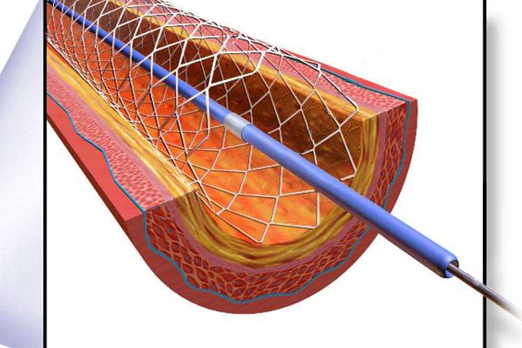
Your doctor usually inserts a stent using a minimally invasive procedure. They will make a small incision and use a catheter to guide specialized tools through your blood vessels to reach the area that needs a stent. This incision is usually in the groin or arm. One of those tools may have a camera on the end to help your doctor guide the stent.
During the procedure, your doctor may also use an imaging technique called an angiogram to help guide the stent through the vessel.
Using the necessary tools, your doctor will locate the broken or blocked vessel and install the stent. Then they will remove the instruments from your body and close the incision.
Any surgical procedure carries risks. Inserting a stent may require accessing arteries of the heart or brain. This leads to an increased risk of adverse effects.
The risks associated with stenting include:
- an allergic reaction to medications or dyes used in the procedure
- breathing problems due to anesthesia or using a stent in the bronchi
- bleeding
- an infection of the vessel
- kidney stones due to using a stent in the ureters
- a re-narrowing of the artery
Rare side effects include strokes and seizures.
If you have bleeding issues, you will need to be evaluated by your doctor. In general, you should discuss these issues with your doctor. They can give you the most current information related to your personal concerns.
How Is A Kidney Stent Removed

Urologists may remove kidney stents in two different ways. Sometimes, they attach a string to the end of the ureter stent. This string later on, comes out from the urethra of patients i.e. the tube, where they urinate. Even string may pull on the ureter stent to remove it. In cases, when urologists do not attach a string, they insert a small camera i.e. a cystoscope in the urethra of patients after administering a suitable numbing medication. Cystoscope thus advanced in the bladder to allow grasping of the stent by the help of respective instrument and later on, removed.
Also Check: Miralax And Kidneys
What Is A Kidney Stent
Rarely, a healthcare provider cant place a ureteral stent due to scarring or other problems. You may need a nephrostomy instead. To perform nephrostomy, a radiologist inserts a stent directly into a kidney. The kidney stent drains urine from the kidney into a bag outside of the body, bypassing the ureters and bladder.
A note from Cleveland Clinic
Ureteral stenting is an effective way to allow painful kidney stones to pass through the ureters and out of the body. Ureteral stents for kidney stones and ureteral stones are temporary. Some people need ureteral stents longer to keep narrowed ureters open. A ureteral stent can be uncomfortable and even slightly painful. Your healthcare provider can suggest ways to ease discomfort until its time to remove the stent.
Last reviewed by a Cleveland Clinic medical professional on 08/25/2021.
References
Time Your Fluid Intake
Youll want to drink plenty of water after you have a stent placed. This will help you flush blood and urine through your kidneys.
However, drinking too much water close to bedtime can cause you to make several additional trips to the bathroom at night.
To address this concern, try to drink plenty of water during the day and start to taper off your intake after dinner. This can help reduce the urinary frequency and urgency you may experience at night.
Your goal will be to keep your urine pale yellow whenever possible. This color indicates that youre hydrated.
Don’t Miss: Are Almonds Bad For Your Kidneys
How Does The Procedure Work
You will lie on your stomach, usually with one side slightly raised on a pillow, and will receive an injection of a painkiller and a sedative. The interventional radiologist will then insert a cystoscope through your urethra into your bladder. This cystoscope will then be used to find the opening where the ureter connects to your bladder. After detecting this opening, the IR will thread the ureteral stent into your ureter via the cystoscope. Once the stent is firmly in place, the IR will remove the cystoscope.
A ureteric stent must be changed every three to six months. This is usually performed as an out-patient procedure.
What To Expect Back Home
At hospital discharge, your doctor or nurse will give you instructions for rest, driving, and doing physical activities after the procedure.
Because surgical instruments were inserted into your urinary tract, you may experience urinary symptoms for some time after surgery. These symptoms usually disappear in a few weeks.
Symptoms may include:
- a mild burning feeling when urinating
- small amounts of blood in the urine
- mild discomfort in the bladder area or kidney area when urinating
- need to urinate more frequently or urgently
- pain resulting from an internal abrasion that needs time to heal
Try to drink fluids often but in small quantities. Sometimes a blood clot can cause pain . The urine contains a substance that will dissolve this clot.
If the pain remains despite the use of pain medication, contact the hospital or your doctor.
Recommended Reading: Is Celery Good For The Kidneys
How Is It Done
Ureteric stenting can be done in two ways:
- It can be done through the kidneys via a nephrostomy
If you need a nephrostomy tube as well as ureteric stenting, your doctor may do both procedures at the same time. It is done by initially performing the nephrostomy and then using the nephrostomy to insert the stent into the ureters. This process is usually done in a radiology department under Ultrasound and x-ray guidance. General anaesthesia is usually not required for this and the procedure can be performed using sedation.
- It can also be done through the urinary bladder using a camera called a cystoscope or ureteroscope. Your doctor will pass a camera through the water passage and will access the urinary bladder. Once in the bladder, the lower opening of ureters can be seen through which the stent can be passed upwards to the kidney. It is usually done under general anaesthetic and using x-ray guidance in an operation theatre.
How Do Doctors Insert A Urinary Stent
When the passage between a person’s kidneys and bladder becomes blocked, a doctor may perform a procedure to insert a urinary stent into the tube between them, the ureter. First, it is necessary to determine which size and type of stent is best suited to the patient’s needs. The procedure is typically performed in the hospital, under general anesthesia, though in some cases the patient is kept awake and given local anesthesia. Placement of the stent is usually accomplished by insertion through the urethra, which is the tube that leads from the bladder out of the body, though in certain cases an incision through the skin may be needed to reach the ureter.
There are a number of different types of urinary stent, and the right one needs to be chosen for a patient before the procedure can be done. Depending on the patient’s needs and physiology, different materials, sizes, and designs of stent might be used. For example, a patient getting a stent put in temporarily may be better suited for an open-ended stent, while someone having one placed more permanently may fare better with one that has coiled ends to hold it in place. A stent may also have a string attached for the doctor to later pull it out, or it may not if the doctor intends to remove it with a cytoscope.
Don’t Miss: Kidney Apple Cider Vinegar
What Are Ureteral Stenting And Nephrostomy
Urine is normally carried from the kidneys to the bladder through long, narrow tubes called ureters. The ureter can become obstructed due to conditions such as kidney stones, tumors, infection, or blood clots. When this happens, physicians can use image guidance to place stents or tubes in the ureter to restore the flow of urine to the bladder.
A ureteral stent is a thin, flexible tube threaded into the ureter. When it is not possible to insert a ureteral stent, a nephrostomy is performed. During this procedure, a tube is placed through the skin on the patient’s back into the kidney. The tube is connected to an external drainage bag or from the kidney to the bladder for internal drainage.
How Is A Ureteral Stent Removed

A ureteral stent that’s going to be in place for only a few days to a week usually has a string attached to the end of it. This string comes out of the urethra and is taped to the child’s leg. This type of stent is removed either at home or in the doctor’s office.
Stents that are in place for several weeks or months are removed by the urologist in the operating room.
You May Like: How Mich Is A Kidney Worth
Side Effects And Complications
The main complications with ureteral stents are dislocation, infection and blockage by encrustation. Recently stents with coatings, such as heparin, were approved to reduce infection and encrustation to reduce the number of stent exchanges.
Other complications can include increased urgency and frequency of urination, blood in the urine, leakage of urine, pain in the kidney, bladder, or groin, and pain in the kidneys during, and for a short time after urination. These effects are generally temporary and disappear with the removal of the stent. Drugs used for the treatment of OAB are sometimes given to reduce or eliminate the increased urgency and frequency of urination caused by the presence of the stent.
Stents often have a thread, used for removal, that passes through the urethra and remains outside the body. This thread may cause irritation of the urethra. This may be increased for patients who were born with Hypospadias or other conditions that required a similar corrective surgery. Care must be taken to ensure that the thread is not caught or pulled, which may dislodge the stent.
Why Is A Stent Used
A stent is most commonly used to bypass an obstruction of the ureter, to allow the ureter to heal after surgery, and to treat kidney stones. A stent is sometimes placed emergently and can be lifesaving when there is an obstruction of the ureter and an associated infection. If this is due to a stone, the stone is left in place and will be treated later due to the infection.
As an added benefit the stent allows the ureter to passively become larger, which can make the future procedure to remove the stone less traumatic and allow any stone fragments to pass more easily once the stent is removed. A stent is also placed at times during stone surgery to allow the ureter to heal or prevent fragments from obstructing the kidney.
It is most commonly inserted by passing a scope with a camera into the urethra and bladder. The stent is then inserted into the opening of the ureter to the kidney using x-rays to visualize its placement.
Read Also: Pomegranate Juice And Kidney Stones
Who Interprets The Results And How Do I Get Them
After the procedure is complete, the interventional radiologist will tell you whether the procedure was a success.
Your interventional radiologist may recommend a follow-up visit.
This visit may include a physical check-up, imaging exam, and blood tests. During your follow-up visit, tell your doctor if you have noticed any side effects or changes.
Why Would I Need A Stent
Stents are usually needed when plaque blocks a blood vessel. Plaque is made of cholesterol and other substances that attach to the walls of a vessel.
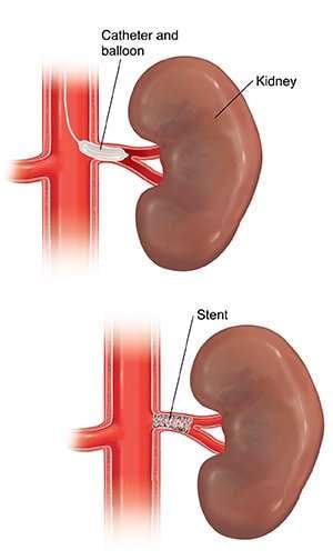
You may need a stent during an emergency procedure. An emergency procedure is more common if an artery of the heart called a coronary artery is blocked. Your doctor will first place a catheter into the blocked coronary artery. This will allow them to do a balloon angioplasty to open the blockage. Theyll then place a stent in the artery to keep the vessel open.
Stents can also be useful to prevent aneurysms from rupturing in your brain, aorta, or other blood vessels.
Besides blood vessels, stents can open any of the following passageways:
- bile ducts, which are tubes that carry bile to and from digestive organs
- bronchi, which are small airways in the lungs
- ureters, which are tubes that carry urine from the kidneys to the bladder
These tubes can become blocked or damaged just like blood vessels can.
Preparing for a stent depends on the type of stent being used. For a stent placed in a blood vessel, youll usually prepare by taking these steps:
Youll receive numbing medicine at the site of the incision. Youll also get intravenous medication to help you relax during the procedure.
You May Like: Can A Kidney Stone Cause Constipation
What Are Ureteral Stents
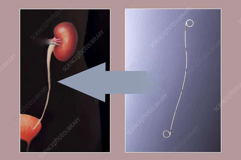
Ureteral stents are thin, flexible tubes that hold ureters open. The ureters are part of the urinary system. Typically, these long, thin tubes carry urine from the kidneys to the bladder. Healthcare providers place ureteral stents to prevent or treat ureteral obstructions.
Silicone or polyurethane ureteral stents are about 10 to 15 inches long and about ¼ inch in diameter. They line the entire length of the ureter, keeping it open. The top part of the stent has a coil that sits inside a kidney. The loop at the lower end sits inside the bladder.
What Will I Experience During And After The Procedure
The doctor or nurse will attach devices to your body to monitor your heart rate and blood pressure.
You will feel a slight pinch when the nurse inserts the needle into your vein for the IV line and when they inject the local anesthetic. Most of the sensation is at the skin incision site. The doctor will numb this area using local anesthetic. You may feel pressure when the doctor inserts the catheter into the vein or artery. However, you will not feel serious discomfort.
If the procedure uses sedation, you will feel relaxed, sleepy, and comfortable. You may or may not remain awake, depending on how deeply you are sedated.
You may feel slight pressure as the catheter is inserted into the kidney and down the ureter. During placement of a ureteral stent, until the stent is positioned, you may feel pressure as the guide wire is inserted into the bladder resulting in a sensation to urinate. You may experience blood-tinged urine for several days following the procedure, which will usually clear up on its own.
You will remain in the recovery room until you are completely awake and ready to return home.
You will not feel when the contrast is excreted into the urine.
You should be able to resume your normal activities within a few days.
Read Also: Is Apple Cider Vinegar Bad For Kidney Disease
How Is A Stent Removed
Stents can be removed two different ways. Sometimes a string has been left attached to the end of the stent. This string is allowed to come out of the patients urethra . The string can be used to pull on the stent and remove it. If a string is not attached, numbing medication is usually administered, and a small camera called a cystoscope is inserted into the urethra. The cystoscope is advanced into the bladder along with a grasping instrument, which grasps and removes the stent.
Ureteral Stenting And Nephrostomy

Ureteral stenting and nephrostomy help restore urine flow through blocked ureters and return the kidney to normal function. Ureters are long, narrow tubes that carry urine from the kidneys to the bladder. They can become obstructed and urine flow blocked as a result of various conditions. Your doctor may use image guidance to place a thin, flexible tube called a stent into the ureter to restore urine flow. If a stent cannot be placed, he may perform a nephrostomy, during which a tube is placed through the skin into the kidney and connected to either an external drainage bag or the bladder for internal drainage.
Tell your doctor if there is a possibility you are pregnant and discuss any recent illnesses, medical conditions, allergies and medications you’re taking. Your doctor may advise you to stop taking aspirin, nonsteroidal anti-inflammatory drugs or blood thinners several days prior to your procedure and instruct you not to eat or drink anything after midnight the night before. Take regular medication with sips of water. Leave jewelry at home and wear loose, comfortable clothing. You may be asked to wear a gown. If you are not to be admitted to the hospital, plan to have someone drive you home afterward.
Recommended Reading: Is Ginger Tea Good For Your Kidneys