Citation Doi And Article Data
Citation:DOI:Dr Yuranga WeerakkodyRevisions:see full revision historySystems:
- Pelviureteric junction stenosis
Pelviureteric junction obstruction/stenosis, also known as ureteropelvic junction obstruction/stenosis, can be one of the causes of obstructive uropathy. It can be congenital or acquired with a congenital pelviureteric junction obstruction being one of the commonest causes of antenatal hydronephrosis.
Anatomy Of Ureteropelvic Junction Obstruction
This term can be better understood if it is broken down a bit. Uretero refers to the ureter. This is the tube that carries urine from the kidney down to the bladder. There is usually one ureter for each kidney.
Pelvic refers to the inner curve of the kidney, which is called the renal pelvis. The point where the ureter attaches to the pelvis of the kidney is thus called the ureteropelvic junction .
Significant UPJ obstruction must be treated because it can cause urine to back up into the kidney. This causes the kidney to swell with urine and potentially become damaged to the point where it can have a loss in some or even all function.
How Is Upj Obstruction Diagnosed
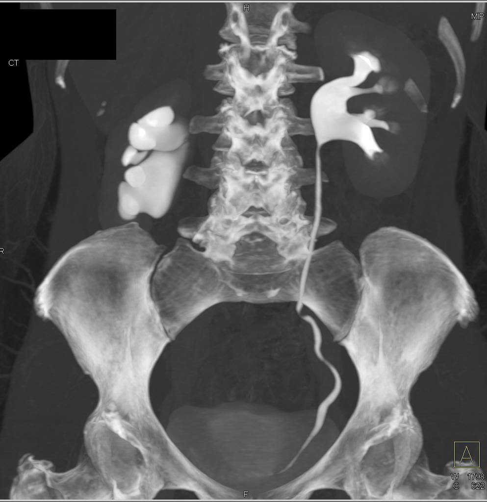
Although ultrasound is a very useful screening test, it is not diagnostic of UPJ obstruction. In order to make the diagnosis, it is necessary to perform a functional test, or one that measures the ability of the kidney to produce and drain urine. The classic examination is called the intravenous pyelogram . In this test, a dye is injected into the bloodstream, and the kidneys remove this substance from the blood. The dye passes into the urine and eventually out of the bladder. The dye is visible on X-ray, and the physician can see the shape of the kidney, renal pelvis and ureter. Although IVPs continue to be helpful, a more useful examination in children is the furosemides renal scan. This test is done in a fashion similar to the excretory urogram except that a radioactive material is used instead of X-ray dye. The material can be followed with a special camera, and this test can give more accurate information about kidney function and drainage.
You May Like: Can You Have 4 Kidneys
What Is Ureteropelvic Junction Obstruction
Ureteropelvic junction obstruction is a blockage in the renal pelvis of the kidney. The renal pelvis is located at the upper end of each ureter . The renal pelvis, which is shaped like a funnel, collects urine.
In normal cases, each of the two kidneys has one ureter. The kidneys filter the blood of waste matter and excess water, creating urine. The urine is pooled at the UPJ, and then flows down the ureters to the bladder.
In UPJ obstruction, the flow of urine is slowed or stopped completely. This raises the risk of kidney damage. In most cases of UPJ obstruction, only one of the kidneys is affected.
What Can Be Expected After Treatment For Upj Obstruction

After repair of UPJ obstruction, there usually is swelling of the ureter and continued poor drainage of the kidney for a period of time. This usually changes as the area heals. The surgeon normally obtains a functional test a few weeks after the procedure to evaluate how well the kidney is working. Patients typically recover quickly from any of the procedures, but some have pain for a few days after surgery. Occasionally, a drainage tube must be left in place to help drain the kidney while it heals. The appearance of the kidney can continue to improve for years, but usually it never looks normal on ultrasound or other studies. Once repaired, a UPJ obstruction almost never recurs. There is nothing that the family can do to prevent further problems with the kidney. Patients may have a slightly increased risk of developing stones and infection throughout their lives because many of the kidneys still contain some pooled urine even though their overall drainage is improved after surgery.
Don’t Miss: Is Wine Bad For Kidney Stones
Will The Kidneys Be Affected Long
Treatment of UPJ obstruction is highly effective. Urologists assess obstructions with nuclear renal scans to determine the best treatment option for each patient. In some cases, the kidney may have to be removed in what is known as a nephrectomy, if the kidney has no function and causes symptoms such as pain, infections, kidney stones.
If you have any questions, to schedule a consultation or if you need a second opinion, pleasecontact us or call:
Dr. Alex Shteynshlyuger is an experienced surgeon who uses advanced techniques individualized for the needs of each patient for treatment of UPJ obstruction including DaVinci Robot, endopyelotomy, and other techniques. Dr. Alex Shteynshlyuger was trained at some of the top hospitals in the United States for Urology. He is experienced in treating some of the most challenging problems in urology.
What Are Ureteropelvic Junction Obstructions
In a normal urinary system, urine flows from the kidney through the ureter and into the bladder. In children with a ureteropelvic junction obstruction, there is a blockage between the ureter and the kidney that can slow or block the flow of urine. In severe cases, the urine is unable to drain from the kidney, and can stretch the organ and cause permanent damage.
- A UPJ obstruction occurs when a blockage between your childs kidney and ureter impedes the flow of urine.
- UPJ obstructions are not very common, occurring in approximately 1 out of every 1,500 babies.
- Many of these blockages are small enough that they wont damage your childs kidney.
- Severe blockages can impair the kidneys ability to drain urine, which can lead to permanent kidney damage.
- If your childs UPJ obstruction is severe enough to put her kidney at risk, a single surgical procedure can be performed to remove the blockage.
Recommended Reading: Can You Have 4 Kidneys
Hydronephrosis And Upj Stones
UPJ stones usually produce significant blockage of the ureter at the UPJ and block urine flow. As a result, urine backs up in the kidney causing dilatation of the renal pelvis and renal calices. This dilatation is called hydronephrosis. Prolonged obstruction of the kidney can cause damage to the kidneys but usually short-term hydronephrosis that lasts less than 1 month causes reversible kidney damage.
Hydronephrosis can also predispose to infection, which is a urological emergency and requires immediate treatment. Usually a ureteral stent or percutaneous nephrostomy is required if a patient with a UPJ stone has a urine infection.
If you have any questions, to schedule a consultation for treatment of kidney stones or if you need a second opinion, pleasecontact us or call:
Dr. Alex Shteynshlyuger is a board certified urologist in NYC who specializes in treating men and women with kidney stones and ureteral stones. He has treated hundreds of men and women with large kidney stones.
- English
Which Treatment Option For Upj Is Best
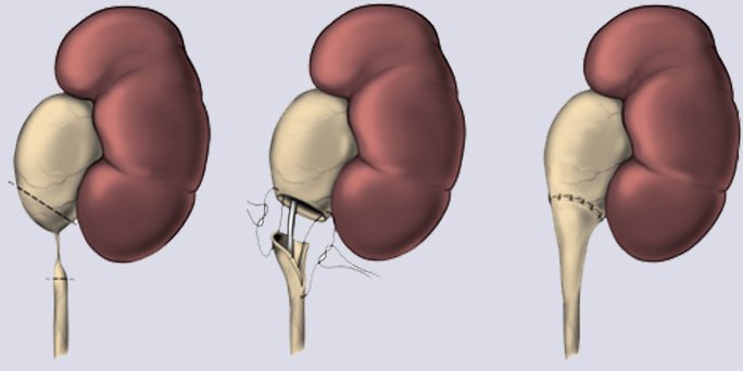
A variety of open surgical procedures have been performed, the most common being the Anderson-Hynes dismembered pyeloplasty, which has a high success rate, as much as 95% and is therefore used as a barometer to compare the utility of less invasive techniques. Pyeloplasty has been performed laparoscopically since 1993. Today, laparoscopic pyeloplasty is most often performed robotically using the DaVinci Robot with success rates that are equivalent to open procedures.
Endourological treatments, such as endopyelotomy have slightly lower long-term success rates and are usually reserved for patients who are not good candidates for laparoscopic repair.
In some patients, typically very elderly who cannot tolerate surgery, an indwelling ureteral stent may be a reasonable treatment option.
The da Vinci robot use for pyeloplasty improved patient outcomes and reduced associated complications.
Don’t Miss: Kidney Pain High Blood Pressure
Imaging Of Crossing Vessels
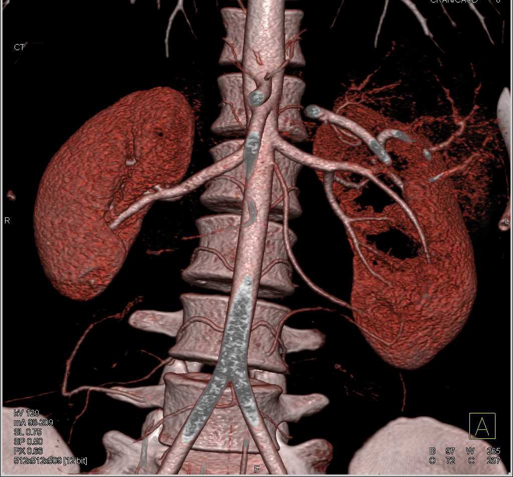
During the past decade, the urologists ability to visualize vessels crossing the UPJ has improved. Earlier attempts to image these vessels relied on intravenous urography to detect abnormalities in the silhouette of the UPJ, thought to be indicative of crossing vessels. One of the signs used most often was the short segment sign, seen as an area of contrast collection between the UPJ obstruction and the crossing vessels. Though a study by Hoffer and Lebowitz showed a 60% sensitivity rate for this sign, attempts to use this as a means of diagnosis by Cassis and coworkers yielded a 20% specificity rate.
A minimally invasive technique used to visualize crossing vessels is helical CT angiography . In a series of 24 consecutive patients with symptomatic UPJ obstruction, 11 of 24 patients were found to have crossing vessels with HCTA visualization. Of those patients, 5 were treated with either laparoscopic or open pyeloplasty, which revealed 100% concordance with the HCTA findings. Farres and colleagues reported similar findings. In this series, 20 patients were examined with HCTA augmented by 3-dimensional reconstruction, and these results were compared with findings noted during open pyeloplasty. Thirteen of the 20 patients were found to have crossing vessels on HCTA, and these findings were confirmed at surgery.
What Causes Ureteropelvic Junction Obstruction
Most UPJ obstructions are present at birth, an indication that structures of the ureter or kidney did not form correctly as the fetus was developing.
In some cases an inherited tendency to obstructions will run in a family, but usually an obstruction appears in just a single family member.
A number of different types of obstructions may be present at birth, such as:
- The opening of the ureter is too narrow.
- There are mistakes in the number or arrangement of small-muscle cells in the ureter. These cells are responsible for the muscular contractions that push urine from the kidney down to the bladder.
- Unusual folds in the walls of the ureter may act as valves.
- Twists may form along the path of the ureter.
- The ureter connects to the renal pelvis in too high a position, creating an abnormal angle between the ureter and kidney.
- An abnormal crossing of blood vessels can press on or distort the UPJ.
Less frequently, UPJ obstructions may form in adults as a result of kidney stones, upper urinary tract infections, surgery, an abnormally crossing blood vessel or swelling in the urinary tract.
Read Also: Cranberry Good For Liver
What Is The Success Rate Of Pyeloplasty Surgery
Open pyeloplasty has a high success rate of 90% in long-term follow-up results . However, it has significant operative morbidities. Laparoscopic pyeloplasty, which was first introduced in 1993 , is minimally invasive, like endourological management, and has a high success rate, like open pyeloplasty .
Treatment Options For Upj Stones Based On Symptoms:

- If severe pain is present along with signs of infection , kidney stones are typically not broken down because that can make things worse. A ureteral double-J stent is placed urgently in the operating room to relieve the obstruction and decompress the kidney. The obstruction is relieved usually with JJ-stent insertion as this is more comfortable for the patient, but percutaneous nephrostomy tube placement is another option, although this is more invasive and a little less convenient for the patient. The urine infection is treated with antibiotics. Once the infection is gone, the stone is then broken with either ureteroscopy and laser or shock-wave, usually in 2-4 weeks. The stone may also be removed with PCNL at a later date once the infection has cleared. The choice of procedure will depend on the size of the stone.
- If pain is present, nausea, and vomiting, but there are no signs of infection, oftentimes the stone can be broken in one sitting such as shock-wave lithotripsy or ureteroscopy with Holmium laser lithotripsy.
- Stones smaller than 8 mm may pass with observation. About 40-60% of stones may pass spontaneously. Stones larger than 7-8 mm that get stuck at UPJ rarely pass without surgical intervention.
Read Also: Does Red Wine Cause Kidney Stones
What Happens Under Normal Conditions
Kidneys produce urine by filtering the blood and removing wastes, salts and water. The urine must then drain from the kidney through an internal collecting system that ends in a funnel-shaped structure called the renal pelvis and into a natural tube called the ureter. Each kidney must have at least one functional ureter to carry the urine from the kidney to the bladder.
How Much Does Urinary Tract Obstruction Cost In Cats
I was told 2400 is more normal for cats who are on the 2 or 3rd obstruction. It depends on the type of surgery being done and other factors of the surgery for perineal urethrostomy costs may be as high as $4,000 or more depending on add ons to the surgery and for cystotomy $2,400 is a reasonable price.
Don’t Miss: Medical Term For Kidneys
How Is Ureteropelvic Junction Obstruction Diagnosed Before And After Birth
- An ultrasound exam before a baby is born can show a UPJ obstruction. As urine gets backed up due to blockage, the kidney swells beyond its normal size, a condition known as hydronephrosis.
- Once the baby is born, tests that measure how well urine is being produced and drained include:
- Blood samples and urine samples such as blood urea nitrogen and creatinine tests provide clues on how well the kidneys are filtering the blood.
- An intravenous pyelogram injects a dye into the bloodstream that is then traced by X-ray as it flows through the kidney, renal pelvis and ureter.
- A nuclear renal scan uses a radioactive substance instead of a dye, and can be traced with a special camera. This shows the functioning of the kidney and how much blockage may be present.
What You Need To Know
Causes of UPJ
- Most commonly an aberrant artery, vein or both called a crossing vessel
- As the blood vessel enlarges with age, it causes the ureter to kink and the renal pelvis to enlarge
Symptoms
- Pain in the back or kidney, especially when passing higher volumes of urine
- Potentially, kidney stones
- Kidney Dilation
Treatment
- Dr. Engel combines traditional laparoscopy with robotic surgery for maximal results
Outcomes
Also Check: Is Pomegranate Juice Good For Your Kidneys
Urinary Reconstruction And Diversion After Cystectomy
Often bladder cancer can require complete bladder removal, and for men, usually means the prostate as well.
For women this can mean removing the bladder plus the uterus, fallopian tubes, ovaries and top of the vaginal wall. When the bladder is removed, the surgeon must reconstruct the bladder so that the urine can pass from the kidney out of the body. The kidneys which make the urine, the ureters which pass the urine to the bladder, and urethra which passes the urine out of the body are all still in place. There is no known artificial bladder has yet, the surgeon must create one one from intestine. Please See the Bladder cancer treatment page for more information.
Treatment Options For Upj Obstruction
Prompt detection and effective treatment strategies for UPJ obstruction are critical to prevent or minimize long-term damage to the kidney. Whereas open surgery procedures comprised first-line treatment strategies in the past, less invasive endo-urological and laparoscopic techniques have recently become popular interventions for UPJ obstruction. For some patients who have symptomatic UPJ obstruction and minimal kidney function, removal of the non-functioning kidney may be a good option.
Read Also: Is Honey Good For Kidney
What Are The Symptoms Of Upj Obstruction
Symptoms of UPJ obstruction may be an abdominal mass a urinary tract infection with fever flank pain, especially with increased fluid intake stones and bloody urine. Patients with UPJ obstruction also may have pain without an infection. Some UPJ obstructions are irregular in nature, and urine may drain normally at one time and be completely obstructed at others, producing sporadic pain.
Treatment Of Upj Obstruction

- If your child is young and is an infant or small child, open surgical correction is generally performed.
- The surgery involves cutting the ureter free from the kidney, clearing the obstructed area, and reattaching it. This is called a pyeloplasty and has a success rate of more than 95%.
- During surgery, a stent may be placed in the ureter to temporarily hold it open.
Recommended Reading: Can Seltzer Water Cause Kidney Stones
What Is A Upj Obstruction
Ureteropelvic junctionobstructionblockage
. Considering this, what causes UPJ obstruction?
Causes. Most often UPJ obstruction is congenital. Though it occurs less often in adults, UPJ obstruction may happen after kidney stones, surgery or upper urinary tract swelling. In UPJ obstruction, the kidney makes urine faster than it can be drained through the renal pelvis into the ureter.
Beside above, how is UPJ obstruction treated? The traditional treatment for ureteropelvic junction obstruction has been open surgery to cut out the area of scarring and re-connect the ureter to the kidney. Over the past several years, newer less invasive treatment options have been developed.
Then, what are the symptoms of UPJ obstruction?
Symptoms of UPJ obstructions include:
- The renal pelvis and/or kidneys are dilated
- Urinary tract infection.
- Poor growth in infants
- Back pain.
Ureteropelvic Junction Obstruction In Adults
A 51-year-old man presents to his primary care physician with complaints of fatigue and lethargy over the past few months. He also notes left flank and inguinal pain, which has become progressively more symptomatic over the past few weeks. He has no prior history of kidney stones or abdominal surgery.
Physical examination was remarkable only for mild left costovertebral angle tenderness. Routine laboratory testing revealed a creatinine level of 2.0 mg/dL, which was increased from his baseline of 1.1 mg/dL from the previous year. Results of a renal ultrasound demonstrated a normal-sized left kidney with moderate hydronephrosis and slight thinning of the parenchyma his right kidney was normal appearing with no renal stones . Laboratory testing was repeated, and his creatinine level remained elevated at 1.7 mg/dL, with an estimated glomerular filtration rate of 43 mL/min/1.73 m2. Because of his compromised renal function, a computed tomography scan was obtained without intravenous contrast. The scan demonstrated moderate left hydronephrosis with slight thinning of the parenchyma and the suggestion of a lower pole-crossing vessel . Overall, the findings appeared to be consistent with a ureteropelvic junction obstruction. A MAG-3 renal scan with furosemide demonstrated a compromised left renal function with delayed uptake, poor cortical transit, minimal excretion, and little response to furosemide. Left renal split function was reported to be 9%.
Recommended Reading: Is Wine Good For Kidney Stones