Characteristics Of Epithelial Layers
Epithelial tissue is composed of cells laid out in sheets with strong cell-to-cell attachments. These protein connections hold the cells together to form a tightly connected layer that is avascular but innervated in nature.
The epithelial cells are nourished by substances diffusing from blood vessels in the underlying connective tissue. One side of the epithelial cell is oriented towards the surface of the tissue, body cavity, or external environment and the other surface is joined to a basement membrane. The basement layer is non-cellular in nature and helps to cement the epithelial tissue to the underlying structures.
Characteristics Of The Epithelial Tissue
The epithelial tissue has a general characteristic that their cells are closely bound to each other through specialized structures such as tight junctions, desmosomes, interdigitations, gap junctions, and intercellular bridges. These gap junctions help in communication with 2 cells together by cementing each other. They constitute the linings of organs and body parts. They have 1 surface that is not attached to any cell, either exposed to the external environment or the lumen of an organ. They do not possess blood vessels and their nutrients requirement is fulfilled by connective tissue forming the basement membrane.
Image: The 3 characteristics of any epithelium. By Lecturio.
S And Types Of Secretion
Exocrine glands can be classified by their mode of secretion and the nature of the substances released, as well as by the structure of the glands and shape of ducts . Merocrine secretion is the most common type of exocrine secretion. The secretions are enclosed in vesicles that move to the apical surface of the cell where the contents are released by exocytosis. For example, watery mucous containing the glycoprotein mucin, a lubricant that offers some pathogen protection is a merocrine secretion. The eccrine glands that produce and secrete sweat are another example.
Figure 5. Modes of Glandular Secretion. In merocrine secretion, the cell remains intact. In apocrine secretion, the apical portion of the cell is released, as well. In holocrine secretion, the cell is destroyed as it releases its product and the cell itself becomes part of the secretion.
Apocrine secretion accumulates near the apical portion of the cell. That portion of the cell and its secretory contents pinch off from the cell and are released. The sweat glands of the armpit are classified as apocrine glands. Both merocrine and apocrine glands continue to produce and secrete their contents with little damage caused to the cell because the nucleus and golgi regions remain intact after secretion.
Also Check: Bleeding Kidney Treatment
About The Lecturio Medical Online Library
Our medical articles are the result of the hard work of our editorial board and our professional authors. Strict editorial standards and an effective quality management system help us to ensure the validity and high relevance of all content. Read more about the editorial team, authors, and our work processes.
Generalized Functions Of Epithelial Tissue

Epithelial tissues provide the bodys first line of protection from physical, chemical, and biological wear and tear. The cells of an epithelium act as gatekeepers of the body controlling permeability and allowing selective transfer of materials across a physical barrier. All substances that enter the body must cross an epithelium. Some epithelia often include structural features that allow the selective transport of molecules and ions across their cell membranes.
Many epithelial cells are capable of secretion and release mucous and specific chemical compounds onto their apical surfaces. The epithelium of the small intestine releases digestive enzymes, for example. Cells lining the respiratory tract secrete mucous that traps incoming microorganisms and particles. A glandular epithelium contains many secretory cells.
Recommended Reading: Can Kidney Stones Make You Constipated
Why Are Kidney Tubules Lined With Simple Cuboidal Epithelium
Simple cuboidal epithelialkidneyskidney tubules
Hereof, what type of epithelium lines the tubules of the kidneys?
Anatomy Tissue Review
| epithelial tissue whose location is on surface of ovaries | |
| simple cuboidal epithelium | epithelial tissue whose location is lining kidney tubules |
| simple cuboidal epithelium | epithelial tissue whose location is lining the ducts of certain glands |
Furthermore, where is the simple cuboidal epithelium found? Simple cuboidal epithelia are found on the surface of ovaries, the lining of nephrons, the walls of the renal tubules, and parts of the eye and thyroid. On these surfaces, the cells perform secretion and absorption.
Likewise, what is the function of simple cuboidal epithelial tissue?
Simple cuboidal epithelium consists of a single layer cells that are as tall as they are wide. The important functions of the simple cuboidal epithelium are secretion and absorption. This epithelial type is found in the small collecting ducts of the kidneys, pancreas, and salivary glands.
What is epithelial tissue state the type of epithelial tissue present in the lining of blood vessels?
Squamous epithelium is found in the lining of the blood vessels.
Which Type Of Epithelial Tissue Forms The Lining Of Kidney Tubules And Ducts Of Salivary Glands Where It Provides Mechanical Support
Which type of epithelial tissue forms the lining of kidney tubules and ducts of salivary glands, where it provides mechanical support?
Epithelial tissue forms a lining all over the body of the organism in order to protect the inner lying parts. They act as a barrier that keep the different body systems separate. Cuboidal epithelial tissue forms the lining of kidney tubules and ducts of salivary glands, where it provides mechanical support.
Recommended Reading: Is Cranberry Juice Good For Your Liver And Kidneys
Slide 210 Kidney H& e View Virtual Slide
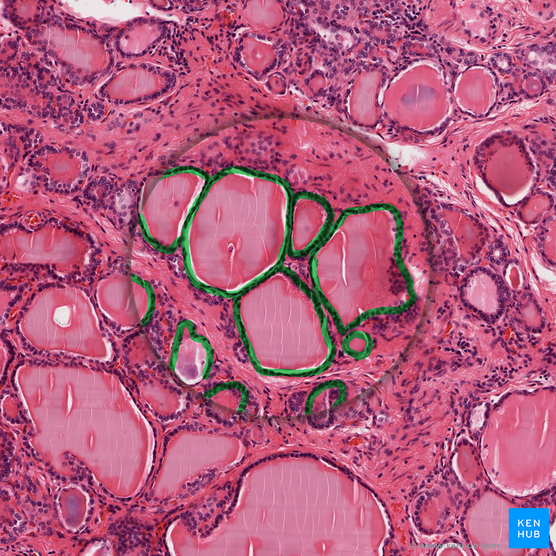
These slides show simple cuboidal epithelium, lining tubules in the kidney. The tubules are cut in all different orientations look for a region toward the middle of the slide where the tubules are cut more or less in longitudinal section in slide 9N-1 View Image or slide 210 View Image and appear as parallel wavy rows . Look for a favorable area where you can see a space lined on either side with simple cuboidal epithelium. Note also that there is very little other tissue between tubules, so that you often see two rows of cuboidal epithelia from adjacent tubules back to back. In other parts of the section, look for tubules in cross-section in slide 9N-1 View Image or slide 210 View Image where the lumen will be surrounded by a circle of cells.
Sensing The Extracellular Environment
“Some epithelial cells are ciliated, especially in respiratory epithelium, and they commonly exist as a sheet of polarised cells forming a tube or tubule with cilia projecting into the lumen.” Primary cilia on epithelial cells provide chemosensation, thermoception, and mechanosensation of the extracellular environment by playing “a sensory role mediating specific signalling cues, including soluble factors in the external cell environment, a secretory role in which a soluble protein is released to have an effect downstream of the fluid flow, and mediation of fluid flow if the cilia are motile.”
Don’t Miss: Renal Diet Orange Juice
Functions Of The Epithelium
Epithelia tissue forms boundaries between different environments, and nearly all substances must pass through the epithelium. In its role as an interface tissue, epithelium accomplishes many functions, including:
Slide 29 View Virtual Slide
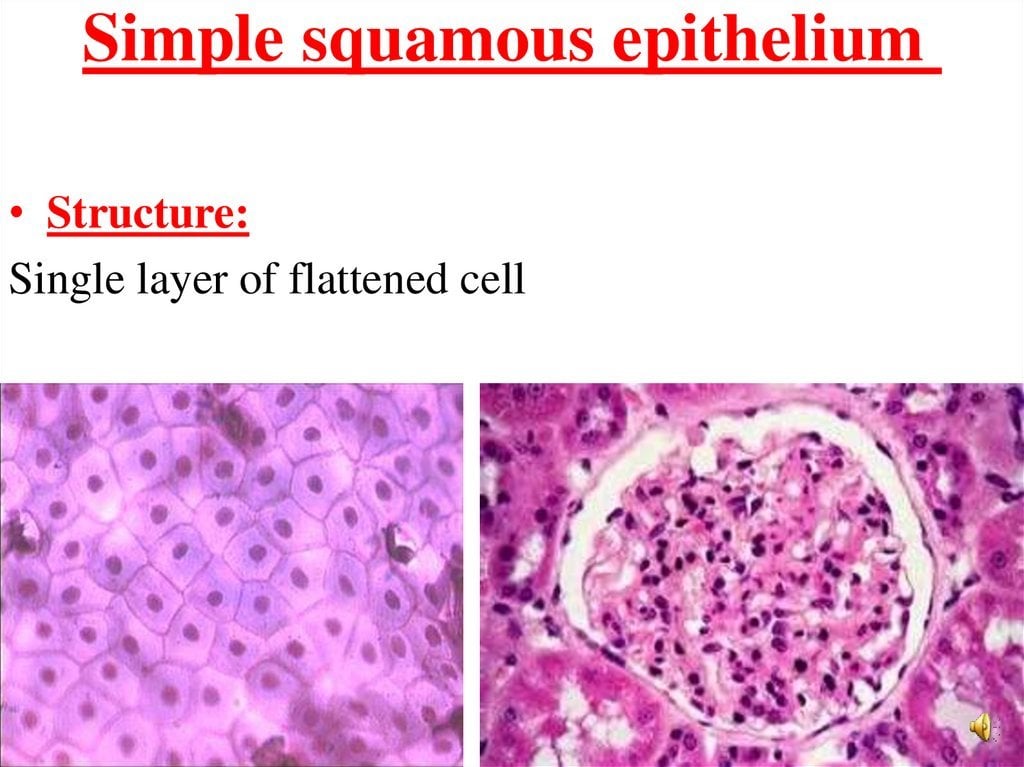
Simple squamous epithelial cells are flattened, i.e., wider than they are tall. A simple squamous epithelium, called “endothelium,” lines blood vessels, lymphatic vessels, and the chambers of the heart. When sections through endothelial cells are viewed with the light microscope, the cytoplasm cannot be seen, because the flattened cell is so thin. Thus, endothelium is generally identified on the basis of the structure and position of nuclei alone that is, the nuclei are also often flattened and elongated, and are found lining the lumen of the vessel. Observe the endothelial lining of blood and lymph vessels in the mesentery in slide 30 View Image. Sometimes the blood vessels contain red blood cells and can be identified that way. Otherwise, look for tubular or circular profiles at low power and examine the endothelial lining of these vessels at high power. Note that the endothelium may be damaged during processing such that it separates from the vessel wall or it may slough off entirely and not be visible at all. In areas where you can find an endothelium, note that the nuclei do not always look flattened in vessels that have contracted. Another excellent place to look for endothelial cells is in the many small vessels in the wall of the intestine shown in slide 29 –look for the vessels in the submucosal layer View Image.
Recommended Reading: Does Red Wine Cause Kidney Stones
The Lining Of Blood Vessels Lung Alveoli And Kidney Tubules Are All Made Up Of Epithelial Tissue
True
Lining of blood vessels, lung alveoli and kidney tubules are all made of squamous and cuboidal epithelial tissues. The lining of the blood vessel is called endothelium and is made of simple squamous epithelial tissue, alveoli are sac like structures made of simple squamous epithelial tissue. The kidney tubules are lined by cuboidal epithelial tissue.
Return To The Histology Tutorial Menu

Epithelial surfaces are plentiful in the human body. The entire body has an epithelial covering called skin. The respiratory tract, gastrointestinal tract, and urinary tract all have epithelial linings. Any glandular, exocrine secretion must pass through an epithelial-lined duct.
Epithelia form a mechanical barrier on surfaces exposed to the external environment. Epithelia can be specialized to perform additional functions such as removing inhaled debris with cilia in the respiratory tract, or absorbing nutrients from the gastrointestinal tract, or secreting mucin and fluid to provide lubrication and transport in ducts.
There are several major types of epithelia:
Read Also: Can You Have 4 Kidneys
Pancreas: A Special Form Of Glandular Tissue
The pancreas has the particular anatomical and physiological characteristics of having both types of glands. Its exocrine portion passes digestive enzymes through the pancreatic duct into the duodenum, while the endocrine portion produces the hormones insulin and glucagon and releases them into the body.
Image: Modes of glandular secretion. By Phil Schatz, License: CC BY 4.0
Browse More Topics Under Tissues
Their functions include protection of the underlying tissues, absorption of substances, regulation of chemicals between the tissues and body cavity etc. And they are able to perform such varied functions since they do not have a definite shape
What is the Connection between Blood and Bones? Learn about Connective Tissue here.
Don’t Miss: Wine For Kidney Stones
Types Of Epithelial Tissue
Epithelial tissues are identified by both the number of layers and the shape of the cells in the upper layers. There are eight basic types of epithelium: six of them are identified based on both the number of cells and their shape two of them are named by the type of cell found in them. Epithelial tissue is classified based on the number of cells, the shape of those cells, and the types of those cells.
| Epithelial Tissue Cells | ||
|---|---|---|
| Function | ||
| Simple squamous epithelium | Air sacs of the lungs and the lining of the heart, blood vessels and lymphatic vessels | Allows materials to pass through by diffusion and filtration, and secretes lubricating substances |
| Simple cuboidal epithelium | In ducts and secretory portions of small glands and in kidney tubules | Secretes and absorbs |
| Simple columnar epithelium | Ciliated tissues including the bronchi, uterine tubes, and uterus smooth are in the digestive tract bladder | Absorbs it also secretes mucous and enzymes. |
| Pseudostratified columnar epithelium | Ciliated tissue lines the trachea and much of the upper respiratory tract | Secrete mucous ciliated tissue moves mucous |
| Stratified squamous epithelium | Lines the esophagus, mouth, and vagina | Protects against abrasion |
| Sweat glands, salivary glands, and mammary glands | Protective tissue | |
| The male urethra and the ducts of some glands. | Secretes and protects | |
| Lines the bladder, urethra and ureters | Allows the urinary organs to expand and stretch |
Stratified Keratinized Squamous Epithelium
Image: Epidermis-structure diagram labeled. By BruceBlaus, License: CC BY 3.0
The outermost cell layers of the epithelium consist of flattened cells with no nuclei and cytoplasm, converting into scales. They are called the stratum corneum, and their purpose is to mechanically protect underlying tissue from dehydration.
The stratified, keratinized squamous epithelium is divided into 5 sections:
- Stratum basale
- Stratum lucidum
- Stratum corneum
The above structure shows an example of the typical skin epitheliumthe epidermis. It is the outermost layer of the 3 layers that make up the skin the inner layers are the dermis and hypodermis.
Recommended Reading: Does Potassium Citrate Reduce Kidney Stones
Viewed From The Surface
Lab-2 28
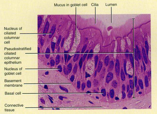
The image to the left shows a model of pseudostratified columnar epithelium. This type of tissue consists of a single layer of cells resting on a noncellular basement membrane that secures the epithelium. The tissue appears stratified because the cells are not all the same height and because their nuclei are located at different levels. Pseudostratified ciliated columnar epithelium lines the trachea and larger respiratory passage ways.
Lab-2 29
Skeletal muscle is the most abundant type of muscle tissue found in the vertebrate body, making up at least 40% of its mass. Although it is often activated by reflexes that function in automatically in response to an outside stimulus, skeletal muscle is also called voluntary muscle because it is the only type subject to conscious control. Because skeletal muscle fibers have obvious bands called striations that can be observed under a microscope, it is also called striated muscle. Note that skeletal muscle cells are multinucleate, that is, each cell has more than one nucleus.
Lab-2 30
Lab-2 31
Cardiac muscle is striated like skeletal muscle but adapted for involuntary, rhythmic contractions like smooth muscle. The myofibrils are transversely striated, but each cell has only one centrally located nucleus. Note the dark blue transverse bands on the model called intercalated disks that mark the boundaries between the ends of the muscle cells. These specialized junctional zones are unique to cardiac muscle.
Lab-2 32
Lab-2 33
Identify The Type Of Tissue In The Following: Skin Bark Of Tree Bone Lining Of Kidney Tubule Vascular Bundle
Answer is skin:Squamous epithelial tissue-
Vascular bundle: Complex permanent tissue –
- Vascular bundles are a collection of tube-like tissues that flow through plants, transporting substances to various parts of the plant.
- Describes a single layer of cells that are flat and scale-like in shape.
The bark of tree: Epidermal tissue/cork-
- It is the outermost layer of the cell that covers the whole plant
Bone: Connective tissue-
- Connective tissue provides support, binds together, and protects tissues and organs of the body.
The lining of kidney tubule: Cuboidal epithelial tissue-
- Simple cuboidal epithelium is a layer of cube-like shape cells, i.e. are as wide as tall.
Also Check: Is Cranberry Juice Good For Your Liver And Kidneys
Fill In The Blanks Lining Of Blood Vessels Is Made Up Of : : : : : : : : Lining Of Small Intestine Is Made Up Of : : : : : : : : Lining Of Kidney Tubules Is Made Up Of : : : : : : : : Epithelial Cells With Cilia Are Found In : : : : : : : : Of Our Body
Fill in the blanks Lining of blood vessels is made up of ________ Lining of small intestine is made up of ________ Lining of kidney tubules is made up of ________ Epithelial cells with cilia are found in ________ of our body.
a) Squamous epitheliumSquamous epithelium are single layer of flat cells and are often permeable. Lining of blood vessels are made up of squamous eithelium.b) Columnar epitheliumColumnar epithelium are uni-layered cells. They line most of the organs of digestive tract.c) Cuboidal epitheliumCuboidal epithelium consists of single layered of cube like cells. They are found in the lining of nephrons .d) Respiratory tractEpithelial cells with cilia occur in our respiratory tract. Cilia move back and forth to help the movement of particles.
A Stratified Squamous Epithelium

This type of epithelium covers surfaces that are subjected to abrasion. The epithelium is constantly replacing itself by division of the basal layer of cells. These cells change morphology as they move toward the surface and are ultimately sloughed off. They are called “stratified” because there are multiple cell layers, and “squamous” because the outermost layer of cells is flattened. There are two subclasses:
1. Stratified squamous nonkeratinizing epithelium
Slide 250 View Virtual Slide
This type of epithelium covers some internal surfaces that are kept moist by mucus or other fluids. Thus, these epithelia do not need to keratinize to avoid desiccation. The lubrication provided by mucus helps to protect against abrasion. Study this type of epithelium in the esophagus and vagina . Again, cell morphology changes from base to apex of the epithelium, the outermost being “squamous” in appearance whereas the basal cells appear more cuboidal or low-columnar. The orientation of the tissue can be confusing because of connective tissue projections that push up into the epithelium. Unlike keratinizing epithelium, nuclei are still present in most surface cells
2. Stratified squamous keratinizing epithelium
Slide 112 (plantar skin, H& E View Virtual Slide
Don’t Miss: Is Pomegranate Juice Good For Your Kidneys
Module : The Tissue Level Of Organization
- Explain the structure and function of epithelial tissue
- Distinguish between tight junctions, anchoring junctions, and gap junctions
- Distinguish between simple epithelia and stratified epithelia, as well as between squamous, cuboidal, and columnar epithelia
- Describe the structure and function of endocrine and exocrine glands and their respective secretions
Most epithelial tissues are essentially large sheets of cells covering all the surfaces of the body exposed to the outside world and lining the outside of organs. Epithelium also forms much of the glandular tissue of the body. Skin is not the only area of the body exposed to the outside. Other areas include the airways, the digestive tract, as well as the urinary and reproductive systems, all of which are lined by an epithelium. Hollow organs and body cavities that do not connect to the exterior of the body, which includes, blood vessels and serous membranes, are lined by endothelium , which is a type of epithelium.
Epithelial cells derive from all three major embryonic layers. The epithelia lining the skin, parts of the mouth and nose, and the anus develop from the ectoderm. Cells lining the airways and most of the digestive system originate in the endoderm. The epithelium that lines vessels in the lymphatic and cardiovascular system derives from the mesoderm and is called an endothelium.