Name The Kind Of Tissue Found In The Living Of The Kidney Tubule
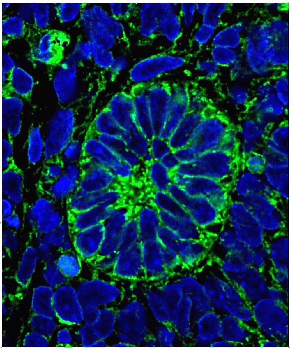
Kidney tubule is made up of epithelial tissues
Simple cuboidal: This tissue is used for the purpose of secretion and absorption. These are also known as epithelial tissues.
I hope the answer is helpful.
Thanks for asking.
The lining of a kidney tubule is made up of Simple Cuboidal Epithelium.
Simple cuboidal epithelium tissues are majorly present in organs which specialized in secretion and perform diffusion such as kidney tubules. These cells are capable of bearing pressure trauma rather than squamous cells. These cells also form the lining of ovaries, linings of nephrons and thyroid.
Kidney Disease Genetic Risk Genes Associated With Distinct Cell Types
Numerous studies have identified causal genes and their variants associated with various kidney diseases10,11. We examined the expression patterns of causal genes involved in chronic kidney disease , albuminuria, IgA nephropathy, nephrolithiasis and lupus nehpritis. Kidney disease-associated genes from GWAS were largely localized in proximal tubule, podocytes, endothelial and myeloid cells. Our analysis confirmed the cell type specific expression of CKD causal gene UMOD in TAL/DCT51, Idiopathic Membranous Nephropathy causal gene PLA2R1 in podocytes52 and eGFRcrea causal gene DACH1 in podocytes53. It also revealed the cell type specific expression of numerous other kidney disease associated genes which were previously unknown – NAT8 in PT cells, IGFBP5 in IC-A and ECs, CUBN in PT cells, and NOTCH4 and DNASE1IL3 in ECs 10,11,54.
Bringing Blood Into An Organoidvascularization Strategies
PSC-derived tissue specific cell types irrespective of being generated in a 2- or 3-dimensional culture are functionally immature despite having morphological properties of the desired cell type. Essential functions of the kidney, including glomerular filtration, tubular reabsorption, or hormone secretion, essentially dependent on the presence of a vasculature. In addition, the diffusion limit of oxygen in organ cultures is around 150 m, while the organoids that are currently being developed range from 1 to 2 mm, raising an issue of oxygen deprivation in the core regions gradually leading to tissue decay.
Read Also: What Does Kidney Failure Pain Feel Like
Build A Kidney: Whats The Blueprint
McMahon led a team of researchers building a blueprint of how the parts fit together and work. The team discovered that timing is critical as the precise arrival of progenitor cells dictates their form and function in the kidney.
Specifically, it takes about 1 million nephrons to form a human kidney. The scientists observed that every time one of these structures forms, the nephron progenitor cells gradually commit to becoming various mature cell types and joining the developing nephron. NPCs that arrive early within the nephron start to differentiate and become the tubule, which controls the reabsorption of important compounds back into the blood and carries urine away. NPCs that occur late develop into the glomerulus, the structure that filters the blood.
Timing is critical in determining the type of mature cell that each progenitor will become.
Nils O. Lindstrom
Timing is critical in determining the type of mature cell that each progenitor will become, said Nils O. Lindstrom, a principal author of the study and a researcher at the stem cell research center, which is part of the Keck School of Medicine of USC.
To show that their predictions were accurate, Lindstrom and colleagues used genetically labeled NPCs in mouse kidneys and grew these under a microscope while capturing images with time-lapse imaging. This allowed them to demonstrate how NPCs gradually move into the newly forming nephron and turn on genes that are specific to particular cell types.
Identify The Type Of Tissue In The Following: Skin Bark Of Tree Bone Lining Of Kidney Tubule Vascular Bundle

Answer is skin:Squamous epithelial tissue-
Vascular bundle: Complex permanent tissue â
- Vascular bundles are a collection of tube-like tissues that flow through plants, transporting substances to various parts of the plant.
- Describes a single layer of cells that are flat and scale-like in shape.
The bark of tree: Epidermal tissue/cork-
- It is the outermost layer of the cell that covers the whole plant
Bone: Connective tissue-
- Connective tissue provides support, binds together, and protects tissues and organs of the body.
The lining of kidney tubule: Cuboidal epithelial tissue-
- Simple cuboidal epithelium is a layer of cube-like shape cells, i.e. are as wide as tall.
Also Check: Is Cranberry Juice Good For Your Liver And Kidneys
Don’t Miss: What Is The Most Common Cause Of Kidney Disease
Integrated Pathway Enrichment Analysis Enables Identification Of Functional Capabilities Of Different Cell Types Of The Kidney
Cell types and subtypes identified by the separated analyses of the sn and sc RNAseq datasets. Bars indicate the percentage of all cells that mapped to a particular cell type or subtype, colors indicate the tissue collection method each particular cell was obtained by. Cell type assignments of separate clusters from sn and sc RNAseq datasets were compared to those obtained by the integrated analysis. Numbers indicate nuclei/cell counts fields are colored by the percentage of cells within each field compared to the row margins. Note that in separated analyses of the sc RNAseq dataset, the applied cutoff for mitochondrial gene expression was higher consequently, some of the cells that were removed in the combined analysis were assigned to cell types in the separated analysis. Similarly, mapping of the nuclei and cells to LMD segments documents that the annotations obtained from the separated analyses map to their correct anatomical origin, as observed for the integrated analysis. All heatmaps are colored according to the number of cells assigned to each LMD subsegment, scaled so each row has mean of 0 and standard deviation of 1. See figure 2A for cell type abbreviations.
Slide 210 Kidney H& E View Virtual Slide
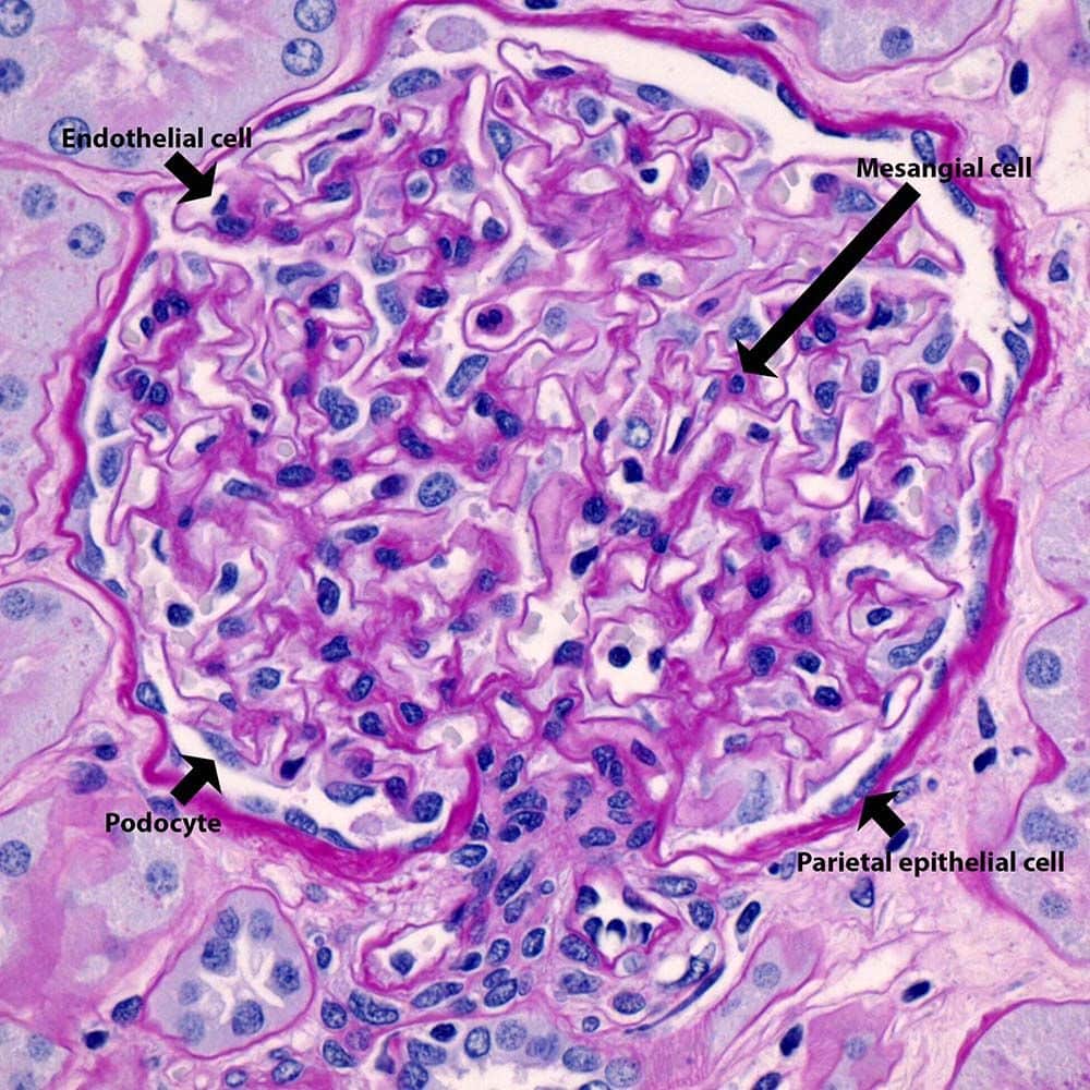
These slides show simple cuboidal epithelium, lining tubules in the kidney. The tubules are cut in all different orientations look for a region toward the middle of the slide where the tubules are cut more or less in longitudinal section in slide 9N-1View Image or slide 210 View Image and appear as parallel wavy rows . Look for a favorable area where you can see a space lined on either side with simple cuboidal epithelium. Note also that there is very little other tissue between tubules, so that you often see two rows of cuboidal epithelia from adjacent tubules back to back. In other parts of the section, look for tubules in cross-section in slide 9N-1 View Image or slide 210View Image where the lumen will be surrounded by a circle of cells.
Don’t Miss: Can Cushing’s Disease Cause Kidney Problems
Xenotransplantation And Blastocyst Complementation
Alternatives to traditional human kidney transplant, such as the xenotransplantation of pig kidneys into humans and blastocyst complementation, have been explored and continue to remain an attractive opportunity to develop more accessible and functional organs for transplant in ESRD patients, Humphreys said. Pfizer first investigated xenotransplantation of pig kidneys into humans in the mid-1990s, but the research was stopped because of concern over porcine endogenous retroviruses , he said. PERV genomes are integrated into the larger genome of a pig and, depending on the class of PERV, can undergo replication in normal pig cells and infect human cells when exposed in culture or via transplant. Unlike other zoonotic pathogens,
Suggested Citation:Exploring the State of the Science in the Field of Regenerative Medicine: Challenges of and Opportunities for Cellular Therapies: Proceedings of a Workshop
PERVs cannot be eliminated through traditional approaches such as biosecure breeding.
Suggested Citation:Exploring the State of the Science in the Field of Regenerative Medicine: Challenges of and Opportunities for Cellular Therapies: Proceedings of a Workshop
rejection remains an issue and that life-long immunosuppression would still be required to maintain tolerance of the transplanted kidney.
Suggested Citation:
Data Generation And Initial Analysis
Seven different RNAseq, proteomics, metabolomics and imaging datasets were generated and analyzed by five different TISes. The PREMIERE TIS generated single cell RNASeq data, the USCD/WashU TIS generated single-nucleus data, the UCSF TIS generated single-cell RNASeq, near-single-cell proteomics and Codex imaging data, the IU/OSU TIS generated laser microcapture dissection RNASeq and LMD proteomics data and the UTHSA-PNNL-EMBL TIS generated spatial metabolomics data.
Read Also: How To Make A Kidney Stone Pass Fast
Kidney’s Maintaining Water Balance
To maintain the blood‘s water balance, the kidneys produce urine which is excreted. This enables the removal of electrolytes, such as sodium and potassium, in excess in the body. Additionally, urine allows the excretion of metabolic waste products from the blood that would otherwise be toxic to the body.
The nephrons maintain water balance in two stages known as the glomerular stage and tubular stage. In the glomerular stage, ultrafiltration occurs whereby glucose, urea, salts and water are filtered at high pressure. Larger molecules, such as proteins and red bloodcells, remain in the blood vessels supplying the kidneys and are filtered out.
Only useful substances are taken back into the blood in the tubular stage. This includes almost all of the glucose, some water and some salts. This ‘purified’ blood returns to circulation.
The substances that have not been reabsorbed travel through the nephron network, to the ureter and to the bladder where it is stored. The urine is then excreted through the urethra. Interestingly, the level of water reabsorption is influenced by the anti-diuretic hormone , which is released from the pituitary gland in the brain. When your body detects low water content in the blood, more ADH is released, which will promote water reabsorption to return your water levels to normal. Read more about this mechanism in our article ADH!
Proteins Specifically Expressed In Distal Tubule
Both the distal tubule and collecting duct are the sites where critical regulatory hormones such as aldosterone and vasopressin regulate acid and potassium excretion and determine the final urinary concentration of K+, Na+, and Cl-. The distal tubule contains the most abundant and most tissue-specific protein in the kidney , although the specific function of this protein is yet somewhat unclear. Similarly, the well-known calbindin is also elevated in the distal tubules. In addition, the list of kidney elevated genes contains several receptors for electrolyte transport, including potassium, sodium, and calcium transporters, such as SLC12A1. Again this is in line with the function of the distal tubule being responsible for the reabsorption of electrolytes to the blood and excretion of potassium to the urine. Another example of a gene expressed in the distal tubule is SLC12A3.
Read Also: How Long Does Kidney Failure Take
Pathway Enrichment Analysis And Module Identification
All Non-glomerular and glomerular metabolites obtained from the three nephrectomy samples were subjected to pathway enrichment analysis using MetaboAnalyst. Some pathways were predicted from metabolites that are general precursors for the synthesis of multiple products and participate in multiple pathways. To exclude such unspecific and consequently uncertain pathway predictions, we focused only on those pathways that were predicted from a pathway specific metabolite . To merge the metabolic pathways with the MBCO SCP-networks, we mapped the MetaboAnalyst pathways âGlycolysis/Gluconeogenesisâ and âGlycerophospholipid metabolismâ to the MBCP SCPs âGlycolysis and Gluconeogenesisâ and to âPhosphoglyceride biosynthesisâ, respectively. Based on identified metabolites, we added the MBCO SCPs âCarnitine shuttleâ and âCarnitine biosynthesis and transportâ to the predicted MetaboAnalyst pathways .
Slide 29 View Virtual Slide

Simple squamous epithelial cells are flattened, i.e., wider than they are tall. A simple squamous epithelium, called âendothelium,â lines blood vessels, lymphatic vessels, and the chambers of the heart. When sections through endothelial cells are viewed with the light microscope, the cytoplasm cannot be seen, because the flattened cell is so thin. Thus, endothelium is generally identified on the basis of the structure and position of nuclei alone that is, the nuclei are also often flattened and elongated, and are found lining the lumen of the vessel. Observe the endothelial lining of blood and lymph vessels in the mesentery in slide 30 View Image. Sometimes the blood vessels contain red blood cells and can be identified that way. Otherwise, look for tubular or circular profiles at low power and examine the endothelial lining of these vessels at high power. Note that the endothelium may be damaged during processing such that it separates from the vessel wall or it may slough off entirely and not be visible at all. In areas where you can find an endothelium, note that the nuclei do not always look flattened in vessels that have contracted. Another excellent place to look for endothelial cells is in the many small vessels in the wall of the intestine shown in slide 29âlook for the vessels in the submucosal layer View Image.
You May Like: Can You Have A Kidney Infection Without A Uti
Important Points Highlighted By Individual Speakers
- Researchers have made strides in using human pluripotent stem cells to build kidney-like organoids that could be used for disease modeling, drug discovery, and toxicity testing for drugs.
- The current treatments for renal diseasedialysis and kidney transplantare expensive and difficult advances in regenerative therapies for renal disease have the potential to make a big difference in patients lives and in the cost of treatment.
- Therapy for polycystic kidney disease should begin much earlier in the course of the disease, meaning that disease detection must improve and that the therapy will need to be safe and tolerable, potentially for decades.
- Blastocyst complementation and xenotransplantation are promising concepts, but they are still very early in the discovery phase. Understanding the scientific basis and complex ethical issues related to both concepts will require years of additional research before they reach the clinic.
There has not been a new treatment for end-stage renal disease developed in nearly 40 years, said Ben Humphreys, the chief of the Division of Nephrology in the Department of Medicine at Washington University School of Medicine in St. Louis. For many patients with kidney disease, the only treatment is dialysis and, potentially, a kidney transplant, but research advances in recent years have generated hope that new therapies based on gene editing, organoids, and even xenotransplantation may one day be available.
Suggested Citation:
Fill In The Blanks Lining Of Blood Vessels Is Made Up Of : : : : : : : : Lining Of Small Intestine Is Made Up Of : : : : : : : : Lining Of Kidney Tubules Is Made Up Of : : : : : : : : Epithelial Cells With Cilia Are Found In : : : : : : : : Of Our Body
Fill in the blanks Lining of blood vessels is made up of ________ Lining of small intestine is made up of ________ Lining of kidney tubules is made up of ________ Epithelial cells with cilia are found in ________ of our body.
a) Squamous epitheliumSquamous epithelium are single layer of flat cells and are often permeable. Lining of blood vessels are made up of squamous eithelium.b) Columnar epitheliumColumnar epithelium are uni-layered cells. They line most of the organs of digestive tract.c) Cuboidal epitheliumCuboidal epithelium consists of single layered of cube like cells. They are found in the lining of nephrons .d) Respiratory tractEpithelial cells with cilia occur in our respiratory tract. Cilia move back and forth to help the movement of particles.
Don’t Miss: How Many Stages Are There To Kidney Disease
S And Types Of Secretion
Exocrine glands can be classified by their mode of secretion and the nature of the substances released, as well as by the structure of the glands and shape of ducts . Merocrine secretion is the most common type of exocrine secretion. The secretions are enclosed in vesicles that move to the apical surface of the cell where the contents are released by exocytosis. For example, watery mucous containing the glycoprotein mucin, a lubricant that offers some pathogen protection is a merocrine secretion. The eccrine glands that produce and secrete sweat are another example.
Figure 5. Modes of Glandular Secretion. In merocrine secretion, the cell remains intact. In apocrine secretion, the apical portion of the cell is released, as well. In holocrine secretion, the cell is destroyed as it releases its product and the cell itself becomes part of the secretion.
Apocrine secretion accumulates near the apical portion of the cell. That portion of the cell and its secretory contents pinch off from the cell and are released. The sweat glands of the armpit are classified as apocrine glands. Both merocrine and apocrine glands continue to produce and secrete their contents with little damage caused to the cell because the nucleus and golgi regions remain intact after secretion.
Also Check: Bleeding Kidney Treatment
A Stratified Squamous Epithelium
This type of epithelium covers surfaces that are subjected to abrasion. The epithelium is constantly replacing itself by division of the basal layer of cells. These cells change morphology as they move toward the surface and are ultimately sloughed off. They are called âstratifiedâ because there are multiple cell layers, and âsquamousâ because the outermost layer of cells is flattened. There are two subclasses:
1. Stratified squamous nonkeratinizing epithelium
Slide 250 View Virtual Slide
This type of epithelium covers some internal surfaces that are kept moist by mucus or other fluids. Thus, these epithelia do not need to keratinize to avoid desiccation. The lubrication provided by mucus helps to protect against abrasion. Study this type of epithelium in the esophagus and . Again, cell morphology changes from base to apex of the epithelium, the outermost being âsquamousâ in appearance whereas the basal cells appear more cuboidal or low-columnar. The orientation of the tissue can be confusing because of connective tissue projections that push up into the epithelium. Unlike keratinizing epithelium, nuclei are still present in most surface cells
2. Stratified squamous keratinizing epithelium
Read Also: How To Get A Kidney Infection