What Toxins Does The Kidney Remove
They rid the body of unwanted products of metabolism such as ammonia, urea, uric acid, creatinine, end products of hemoglobin metabolism, and hormone metabolites toxins that have been made water soluble by phase 2 in the liver and direct excretion of industrial toxins, such as heavy metals and a number of new-to-
What Affects The Amount Of Urine You Produce
The amount of urine you produce depends on many factors, such as the amount of liquid and food you consume and the amount of fluid you lose through sweating and breathing. Certain medicines, medical conditions, and types of food can also affect the amount of urine you produce. Children produce less urine than adults.
Who Can Help Me With A Urinary Problem
Your primary doctor can help you with some urinary problems. Your pediatrician may be able to treat some of your childs urinary problems. But some problems may require the attention of a urologist, a doctor who specializes in treating problems of the urinary system and the male reproductive system. A gynecologist is a doctor who specializes in the female reproductive system and may be able to help with some urinary problems. A urogynecologist is a gynecologist who specializes in the female urinary system. A nephrologist specializes in treating diseases of the kidney.
Don’t Miss: Seltzer And Kidney Stones
Blood Supply And Lymphatics
The ureters receive their blood supply from multiple arterial branches. In the upper or abdominal ureter, the arterial branches stem from the renal and gonadal artery, abdominal aorta, and common iliac arteries. In the pelvic and distal ureter, the arterial branches come from the vesical and uterine arteries, which are branches of the internal iliac artery. The arterial supply will course along the ureter longitudinally creating a plexus of anastomosing vessels. This is of clinical significance because it allows for safe mobilization of the ureter during surgery when proper exposure from surrounding structures is crucial.
The venous and lymphatic drainage of the ureter mirrors that of the arterial supply. The lymphatic drainage is to the internal, external, and common iliac nodes. The lymphatic drainage of the left ureter is primarily to the left para-aortic lymph nodes while the drainage of the right ureter primarily drains to the right paracaval and interaortocaval lymph nodes.
Can Kidney Disease Be Prevented

Yes, in most cases, kidney disease can be prevented. The most common causes of kidney disease are diabetes and high blood pressure. Working with your doctor to prevent these problems, or manage them if you have them, can help prevent kidney disease.
If you already have chronic kidney disease , meaning your kidneys are damaged and cant work as well as they should, you may still be able to prevent kidney failure, which is when your kidneys dont work at all. Following a kidney-friendly diet, being active each day, and understanding your risk factors can help you prevent kidney failure.
Recommended Reading: Kidney Pain High Blood Pressure
Treatment For Urinary Reflux
Most children who have urinary reflux dont need surgery, but may require regular appointments with their doctor.
Children who may need surgery include those who:
- continue to get urinary tract infections while they are on antibiotics
- have other complex abnormalities of the urinary tract.
Surgical correction of reflux consists of either re-inserting the ureters back into the bladder to make a new tunnel, or injecting special material around the bottom of the ureters. Both of these operations restore a functional backflow valve and successfully prevent reflux.
If your child needs surgery, your doctor will discuss the options with you.
What Are The Parts Of The Urinary Tract
People usually have two kidneys, but can live a normal, healthy life with just one. The kidneys are under the ribcage in the back, one on each side. Each adult kidney is about the size of a fist.
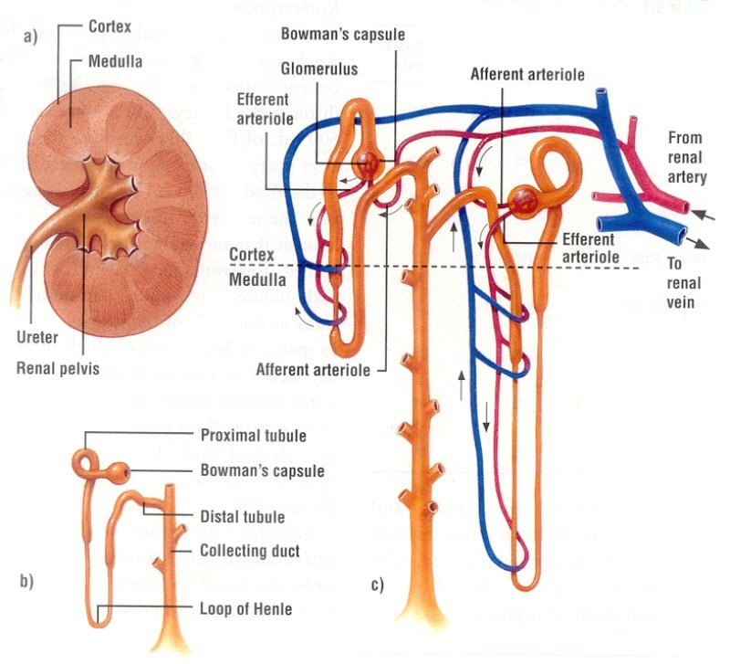
Each kidney has an outer layer called the cortex, which contains filtering units. The center part of the kidney, the medulla , has fan-shaped structures called pyramids. These drain urine into cup-shaped tubes called calyxes .
From the calyxes, pee travels out of the kidneys through the ureters to be stored in the bladder . When a person urinates, the pee exits the bladder and goes out of the body through the urethra , another tube-like structure. The male urethra ends at the tip of the penis the female urethra ends just above the vaginal opening.
Recommended Reading: Does Red Wine Cause Kidney Stones
Structure The Kidneys Are Bean
Gerotas fascia is a thin, fibrous tissue on the outside of the kidney. Below Gerotas fascia is a layer of fat.
The renal capsule is a layer of fibrous tissue that surrounds the body of the kidney inside the layer of fat.
The cortex is the tissue just under the renal capsule.
The medulla is the inner part of the kidney.
The renal pelvis is a hollow area in the centre of each kidney where urine collects.
The renal artery brings blood to the kidney.
The renal vein takes blood back to the body after it has passed through the kidney.
The renal hilum is the area where the renal artery, renal vein and ureter enter the kidney.
The nephrons are the millions of small tubes inside each kidney. Each nephron has 2 parts. Tubules are tiny tubes that collect the waste materials and chemicals from the blood moving through the kidney. The corpuscles contain a clump of tiny blood vessels called glomeruli that filter the blood as it moves through the kidney. The waste products are passed through the tubules to the collecting ducts, which drain into the renal pelvis.
How Are Problems In The Urinary System Detected
Urinalysis is a test that studies the content of urine for abnormal substances such as protein or signs of infection. This test involves urinating into a special container and leaving the sample to be studied. Urodynamic tests evaluate the storage of urine in the bladder and the flow of urine from the bladder through the urethra. Your doctor may want to do a urodynamic test if you are having symptoms that suggest problems with the muscles or nerves of your lower urinary system and pelvisureters, bladder, urethra, and sphincter muscles. Urodynamic tests measure the contraction of the bladder muscle as it fills and empties. The test is done by inserting a small tube called a catheter through your urethra into your bladder to fill it either with water or a gas. Another small tube is inserted into your rectum or vagina to measure the pressure put on your bladder when you strain or cough. Other bladder tests use x-ray dye instead of water so that x-ray pictures can be taken when the bladder fills and empties to detect any abnormalities in the shape and function of the bladder. These tests take about an hour.
Also Check: Can Apple Cider Vinegar Hurt Your Kidneys
What Are The Parts Of The Urinary System
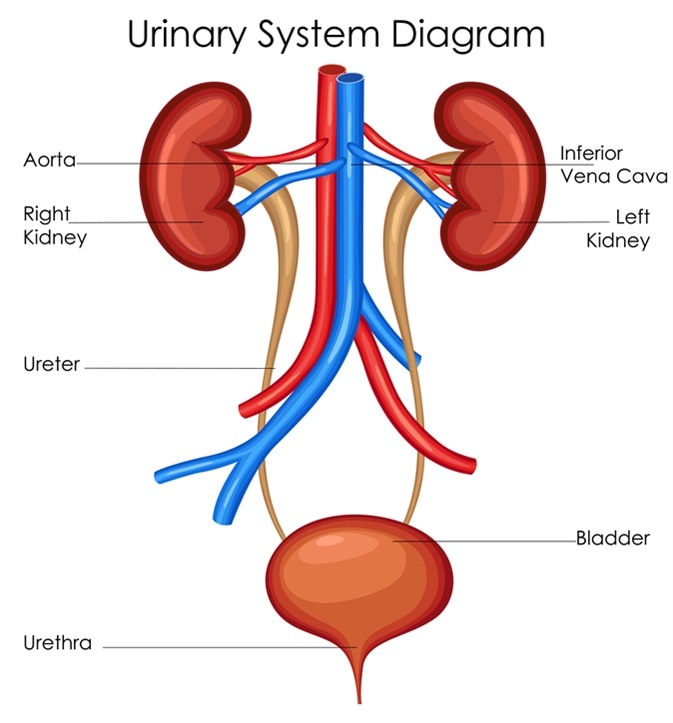
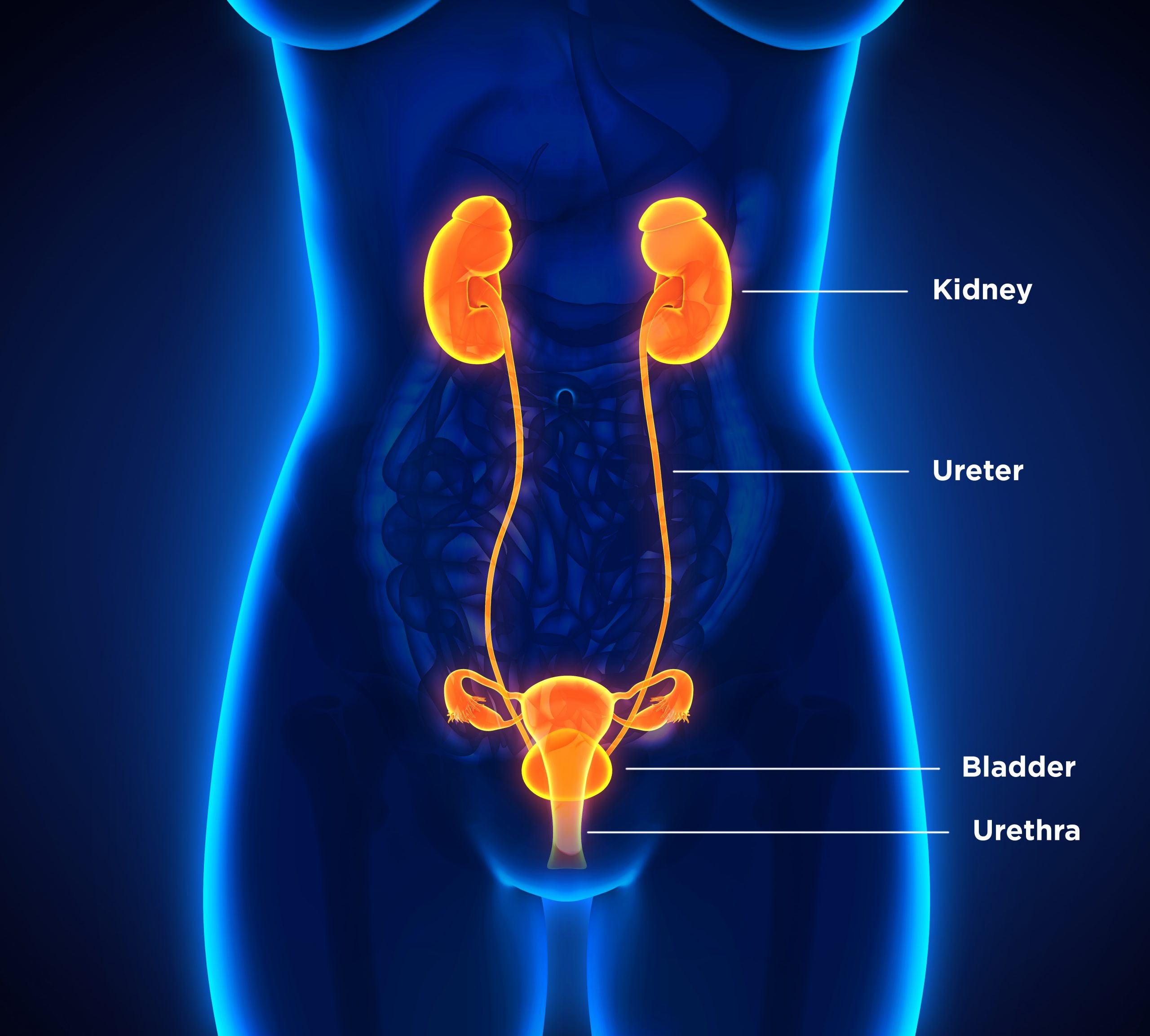
The kidneys, ureters, bladder and urethra make up the urinary system. They all work together to filter, store and remove liquid waste from your body. Heres what each organ does:
- Kidneys: These organs work constantly. They filter your blood and make urine, which your body eliminates. You have two kidneys, one on either side of the back of your abdomen, just below your rib cage. Each kidney is about as big as your fist.
- Ureters: These two thin tubes inside your pelvis carry urine from your kidneys to your bladder.
- Bladder: Your bladder holds urine until youre ready to empty it . Its hollow, made of muscle, and shaped like a balloon. Your bladder expands as it fills up. Most bladders can hold up to 2 cups of urine.
- Urethra: This tube carries urine from your bladder out of your body. It ends in an opening to the outside of your body in the penis or in front of the vagina .
How Does Urination Occur
To urinate, your brain signals the sphincters to relax. Then it signals the muscular bladder wall to tighten, squeezing urine through the urethra and out of your bladder.
How often you need to urinate depends on how quickly your kidneys produce the urine that fills the bladder and how much urine your bladder can comfortably hold. The muscles of your bladder wall remain relaxed while the bladder fills with urine, and the sphincter muscles remain contracted to keep urine in the bladder. As your bladder fills up, signals sent to your brain tell you to find a toilet soon.
Recommended Reading: What Std Messes With Your Kidneys
Female Urology And External Sexual Anatomy
In both men and women, the urology system is the part of the body that deals with urination. It doesn’t take a doctor to know that the urology-related anatomy of men and women look very different, at least from the outside. However, internally, they are similarthe kidneys of both men and women, for example, look and function the same for both genders. But we also differ in some ways, toowomen have much shorter urethras and therefore are at greater risk of bladder infections.
What Is The Urinary Tract

The urinary tract is the bodys drainage system for removing urine, which is made up of wastes and extra fluid. For normal urination to occur, all body parts in the urinary tract need to work together, and in the correct order.
The urinary tract includes two kidneys, two ureters, a bladder, and a urethra.
Kidneys. Two bean-shaped organs, each about the size of a fist. They are located just below your rib cage, one on each side of your spine. Every day, your kidneys filter about 120 to 150 quarts of blood to remove wastes and balance fluids. This process produces about 1 to 2 quarts of urine per day.
Ureters. Thin tubes of muscle that connect your kidneys to your bladder and carry urine to the bladder.
Bladder. A hollow, muscular, balloon-shaped organ that expands as it fills with urine. The bladder sits in your pelvis between your hip bones. A normal bladder acts like a reservoir. It can hold 1.5 to 2 cups of urine. Although you do not control how your kidneys function, you can control when to empty your bladder. Bladder emptying is known as urination.
Urethra. A tube located at the bottom of the bladder that allows urine to exit the body during urination.
The urinary tract includes two sets of muscles that work together as a sphincter, closing off the urethra to keep urine in the bladder between your trips to the bathroom.
Don’t Miss: What Std Messes With Your Kidneys
What Do The Ureter And Urethra Do
The ureter and urethra are both important parts of the body’s urinary tract system. Urine passes from the kidney through the ureter to the bladder. Then, it passes through the urethra to exit the body.
Sometimes, stones, blockages, cancer and other conditions create problems with this part of the urinary tract. Our experts get to the source of the condition that’s making it hard for urine to leave the body successfully and painlessly.
Causes Of Urinary Reflux
Some of the conditions that may cause or contribute to urinary reflux include:
- physical problems of the kidney, present at birth
- physical problems of the bladder and the bladder outlet
- bladder stones
- trauma or injury to the bladder
- temporary swelling after surgery .
A family history of urinary reflux can indicate that someone may be at higher risk of developing urinary reflux.
Don’t Miss: Red Wine Kidney Stones
How Do The Kidneys And Urinary Tract Work
Blood travels to each kidney through the renal artery. The artery enters the kidney at the hilus , the indentation in middle of the kidney that gives it its bean shape. The artery then branches so blood can get to the nephrons 1 million tiny filtering units in each kidney that remove the harmful substances from the blood.
Each of the nephrons contain a filter called the glomerulus . The fluid that is filtered out from the blood then travels down a tiny tube-like structure called a tubule . The tubule adjusts the level of salts, water, and wastes that will leave the body in the urine. Filtered blood leaves the kidney through the renal vein and flows back to the heart.
Pee leaves the kidneys and travels through the ureters to the bladder. The bladder expands as it fills. When the bladder is full, nerve endings in its wall send messages to the brain. When a person needs to pee, the bladder walls tighten and a ring-like muscle that guards the exit from the bladder to the urethra, called the sphincter , relaxes. This lets pee go into the urethra and out of the body.
Does Urethritis Make You Pee A Lot
Urethritis is infection of the urethra, the tube that carries urine from the bladder out of the body. Bacteria, including those that are sexually transmitted, are the most common cause of urethritis. Symptoms include pain while urinating, a frequent or urgent need to urinate, and sometimes a discharge.
You May Like: Fluid Buildup Around Kidney
What Do My Kidneys Do
Every day, your kidneys filter about 30 gallons of blood to remove about half a gallon of extra water and waste products. This waste and extra water make up your urine . The waste comes from the food you eat and the use of your muscles. Your urine travels to your bladder through the ureters, tubes that connect your kidney to your bladder. Your bladder stores the urine until you are ready to urinate . When you urinate, urine leaves your body through your urethra.
Your kidneys also do many other jobs, such as help:
- Make red blood cells
When your kidneys dont work the way they should, they allow waste and water to flow back into your blood stream instead of sending them out with your urine. This causes waste and water to build up in your body, which can cause problems with your heart, lungs, blood, and bones.
Promoting Good Bladder Health
Sometimes, there is no choice but to hold urine, but it may not be good for the bladder. “Holding your urine for a short period of time, usually up to one hour, is typically okay,” Ramin said. “However, protracted and repeated holding of urine may cause over-expansion of bladder capacity, transmission of excess pressure into the kidneys, and the inability to completely empty the bladder. These problems in turn may lead to UTI , cystitis and deterioration of kidney function.”
Drinking plenty of water throughout the day can also help prevent bladder stones by preventing the concentration of minerals that cause the stones. The Mayo Clinic suggests asking a medical profession about how much water the body needs according to age, size and activity level.
Editor’s Note: If you’d like more information on this topic, we recommend the following book:
You May Like: Is Club Soda Good For Kidney Stones
How Does The Urinary System Work
The urinary system’s function is to filter blood and create urine as a waste by-product. The organs of the urinary system include the kidneys, renal pelvis, ureters, bladder and urethra.
The body takes nutrients from food and converts them to energy. After the body has taken the food components that it needs, waste products are left behind in the bowel and in the blood.
The kidney and urinary systems help the body to eliminate liquid waste called urea, and to keep chemicals, such as potassium and sodium, and water in balance. Urea is produced when foods containing protein, such as meat, poultry, and certain vegetables, are broken down in the body. Urea is carried in the bloodstream to the kidneys, where it is removed along with water and other wastes in the form of urine.
Other important functions of the kidneys include blood pressure regulation and the production of erythropoietin, which controls red blood cell production in the bone marrow. Kidneys also regulate the acid-base balance and conserve fluids.
What Is The Path Of Urine Through The Urinary System

From the kidneys through the ureters to the bladder from there through the urethra to be expelled from the body.
Explanation:
Urine is formed after a process of glomerular filtration in the kidneys.
This urine is then conducted through the ureters, twin muscular tubes that connect the kidneys to the bladder, a storage chamber.
The bladder is a muscular chamber that expands as urine fills it.
From the bladder, a muscular tube, the urethra connects to the outside.
The urethra , an internal sphincter at the junction of the urethra and bladder, and an external sphincter comprising the pelvic floor muscles, keep the urine in the bladder till it is ready to expel the urine.
For urination, the bladder walls contract and the urethra and sphincter muscles relax, to allow urine to flow out from the urethra.
Urethra
Explanation:
Urine is formed by thousands of nephrons present inside the paired kidneys and it is passed down the ureters, there from to the urinary bladder. Now how is it formed?
Urine is formed when the blood reaches the malpighian corpuscle that is composed of the Bowman’s capsule and glomerulus. Here most of the blood plasma is filtered out into the Bowman’s capsule.
Glomerular filtrate is taken down the proximal convoluted tubule . Most of the water, glucose and amino acids are reabsorbed here. Active as well as passive reabsorption occurs here.
The resulting fluid passes down the loop of Henle . Electrolytes like Na+ and K+ are reabsorbed here.
Recommended Reading: Is Honey Good For Kidney Health
Renal Pelvis And Ureter
Numerous collecting ducts merge into the renal pelvis, which then becomes the ureter. The ureter is a muscular tube, composed of an inner longitudinal layer and an outer circular layer. The lumen of the ureter is covered by transitional epithelium . Recall from the Laboratory on Epithelia that the transitional epithelium is unique to the conducting passages of the urinary system. Its ability to stretch allows the dilation of the conducting passages when necessary. The ureter connects the kidney and the urinary bladder.