What Is The Function Of The Kidneys
The excess waste products and excess fluid are removed when the kidneys produce urine that is excreted from the body. Moreover, the kidneys play an important role in the regulation of the body’s salt, potassium, and acid content.
The kidneys also produce hormones that stimulate the production of red blood cells that help regulate blood pressure and help control calcium and electrolyte metabolism in the body.
Symptoms Of Kidney Disease
Kidney disease is called a silent disease as there are often no warning signs. People may lose up to 90 per cent of their kidney function before getting any symptoms. The first signs of kidney disease may be general and can include:
- high blood pressure
- changes in the amount and number of times urine is passed
- changes in the appearance of urine
- blood in the urine
- puffiness of the legs and ankles
- pain in the kidney area
- tiredness
- have a family history of kidney failure
- have a history of acute kidney injury
- are of Aboriginal or Torres Strait Islander origin.
Clinical Relevance: Variation In Arterial Supply To The Kidney
The kidneys present a great variety in arterial supply these variations may be explained by the ascending course of the kidney in the retroperitoneal space, from the original embryological site of formation to the final destination . During this course, the kidneys are supplied by consecutive branches of the iliac vessels and the aorta.
Usually the lower branches become atrophic and vanish while new, higher ones supply the kidney during its ascent. Accessory arteries are common . An accessory artery is any supernumerary artery that reaches the kidney. If a supernumerary artery does not enter the kidney through the hilum, it is called aberrant.
Recommended Reading: Does Pop Cause Kidney Stones
Why You Get Stones
Part of preventing stones is finding out why you get them. Your health care provider will perform tests to find out what is causing this. After finding out why you get stones, your health care provider will give you tips to help stop them from coming back.
Some of the tests he or she may do are listed below.
Medical and Dietary History
Your health care provider will ask questions about your personal and family medical history. He or she may ask if:
- Have you had more than one stone before?
- Has anyone in your family had stones?
- Do you have a medical condition that may increase your chance of having stones, like frequent diarrhea, gout or diabetes?
Knowing your eating habits is also helpful. You may be eating foods that are known to raise the risk of stones. You may also be eating too few foods that protect against stones or not drinking enough fluids.
Understanding your medical, family and dietary history helps your health care provider find out how likely you are to form more stones.
Blood and Urine Tests
Imaging Tests
When a health care provider sees you for the first time and you have had stones before, he or she may want to see recent X-rays or order a new X-ray. They will do this to see if there are any stones in your urinary tract. Imaging tests may be repeated over time to check for stone growth. You may also need this test if you are having pain, hematuria or recurrent infections.
Stone Analysis
Imaging Techniques For The Kidney

KUB is the proper terminology for a radiograph of the abdomen when used to view the urinary tract. The outline of kidneys can usually be seen. Ureters usually are not visible. The most common pathological findings are urinary tract stones. See the image below.
The imaging technique of choice for evaluation of the urinary tract and adrenal glands is CT scanning. It allows evaluation of the relative density of structures. CT scanning without contrast can be used for detection of renal or ureteral stones. See the image below.
The advantages of ultrasonography include that it is readily available, does not require contrast, and avoids radiation exposure. The renal medulla is hypoechoic compared with the renal cortex. The renal cortex is isoechoic or slightly hypoechoic compared with the liver. Ultrasonography is able to identify simple or mildly complicated cysts and is able to differentiate these lesions from a solid mass. It is excellent for detecting hydronephrosis. See the image below.
Radionuclide Renal Scintigraphy
Renal radionuclide imaging is an integral part of nuclear medicine and provides substantial information on the actual renal function.
The following radionuclides are used for dynamic imaging:
- Tc-99m-diethylene triamine pentaacetic acid
- Tc-99m-MAG3
For static imaging, Tc-99m-dimercaptosuccinic acid is used.
A diuretic challenge can also be administered.
You May Like: Is Cranberry Juice Good For Your Liver And Kidneys
Kidney Pain Definition And Facts
- The function and purpose of the kidneys are to remove excess fluid and waste products from the body.
- The kidneys are organs that are located in the upper abdominal area against the back muscles on both the left and right side of the body.
- Kidney pain and back pain can be difficult to distinguish, but kidney pain is usually deeper and higher in the and back located under the ribs while the muscle pain with common back injury tends to be lower in the back.
- Common causes of kidney pain are mainly urinary tract infections, kidney infections, and kidney stones. However, there are many other causes of kidney pain, including penetrating and blunt trauma that can result in a “lacerated kidney.”
- If a woman is pregnant and has kidney pain, she should contact her doctor.
- Symptoms of kidney pain may include
- vomiting.
B Decreased Distal Delivery Of Sodium
Mild to moderate reductions in renal perfusion do not typically cause distal delivery of Na+ to fall to a level that impairs K+ secretion sufficiently to result in clinically significant hyperkalemia. In untreated congestive heart failure S is typically normal or high normal despite the reduction in distal Na+ delivery as long as the impairment in cardiac function and renal perfusion is not severe. When such patients are treated with ACEIs or ARBs the fall in circulating aldosterone concentration is typically counterbalanced by increased distal Na+ delivery so that the S remains stable. The increase in distal Na+ is due to the afterload-reducing effects of these drugs, causing an improvement in cardiac output and renal perfusion.
When renal perfusion becomes more severely reduced, as in patients with intractable congestive heart failure, proximal reabsorption can become so intense that very little Na+ escapes into the distal nephron. A lack of Na+ availability can begin to impair renal K+ secretion, particularly in the setting of CKD, where baseline aldosterone levels are often reduced and the capacity for increased production is limited.
Recommended Reading: Can You Have 4 Kidneys
Feeling Tired Or Sluggish During The Day
Everyone has a day when they feel tired maybe you didnt get enough sleep, or ate the wrong foods, or some other temporary factors are at play. But sometimes, fatigue is caused by lack of a hormone called erythropoietin, or EPO. The main function of EPO is to stimulate the production of red blood cells, and red blood cells carry energizing oxygen to cells throughout your body.
Stressed kidneys do not produce enough EPO, thereby reducing the number of red blood cells and making you feel weak and tired out.
Other Types Of Kidney Failure In Cirrhosis
Drug Toxicity
As stated previously, kidney perfusion and GFR in patients with cirrhosis and ascites are maintained by an increased renal synthesis of vasodilating prostaglandins . Nonsteroidal antiinflammatory drugs inhibit prostaglandin synthesis and cause a profound reduction in renal blood flow, with resultant kidney failure occurring in a high proportion of these patients. Patients with cirrhosis and ascites are also susceptible to other nephrotoxins, including aminoglycoside antibiotics and intravenous contrast.
Intravascular Volume Losses
In patients with cirrhosis and upper gastrointestinal bleeding, the incidence of kidney failure is 11%. Risk factors include severity of blood losses and degree of liver failure . A substantial number of patients with kidney failure following bleeding episodes recover kidney function following volume repletion, consistent with a prerenal state. In other patients, however, kidney failure persists or progresses despite resolution of the bleeding episode, suggesting tubular damage or HRS.
Kidney Failure Associated with Infection
Parenchymal Kidney Diseases
Abdominal Compartment Syndrome
Read Also: Does Red Wine Cause Kidney Stones
Crossing Vessel Upj Obstruction Vesicoureteral Reflux
Crossing vessel, ureteropelvic junction obstruction, or vesicoureteral reflux can become pathophysiologic if it causes extrinsic or primary intrinsic obstruction leading to hydronephrosis. This can be seen with aberrant crossing vessels in a single system, which leads to UPJ obstruction. Obstruction can also occur from an ectopic ureter, where it is commonly seen inserting inferomedially in an abnormal location and is often associated with the upper pole moiety of a complete duplicated collecting system.
Similarly, a ureterocele in a single system, or sometimes seen in a complete duplicated system, can cause obstruction. From an intrinsic standpoint, UPJO can also be caused by an adynamic/aperistaltic segment of ureter that is due to abnormal embryologic development. Secondary etiologies of obstruction include stones, infections, iatrogenic ureteral damage causing strictures, and other acquired factors that are not due to anatomic variants.
Vesicoureteral reflux is another variation and is caused by an abnormal insertion of the ureter in the bladder in an abnormal position . This insertion site leads to a shorter intramural tunnel length for the ureter to pass through the bladder wall, which leads to inadequate compression of the ureter during bladder filling and contraction and may allow reflux of urine up the ureter. Vesicoureteral reflux can contribute to pyelonephritis and, in extreme situations, irreversible damage to an affected renal unit.
The Nutcracker Syndrome
Why Lockdowns Have Left Kidney Patients Totally And Completely Terrified
Few need COVID-19 surges to end quite like the 37 million Americans who live with chronic kidney disease.
Nichole Jefferson has been social distancing for more than a decade. The 48-year-old Dallas resident had her first kidney transplant in 2008, and her anti-rejection medications weakened her immune system and left her susceptible to serious infection. So she was experienced with wearing disposable gloves to pump gas and use the ATM, wiping down her groceries, and donning a face mask well before COVID-19 emerged.
Two years ago, her kidney began to fail, and she went back on the transplant list. In April, Jefferson received her second transplant and hoped the surgery would give her more energy to engage with life. Instead, because of the coronavirus crisis, shes more isolated than ever before.
I was totally and completely terrified, Jefferson says.
Jefferson is one of millions of Americans with chronic kidney disease who are finding it hard to cope during the pandemic. Not only are they at higher risk of serious complications from COVID-19, many are also managing the combined financial blows of the economic downturn and their need for high-priced, hard-to-get medications. And COVID-19 appears to be making this worse, as some patients with the virus are developing a rapid version of kidney failure.
For people who are in kidney failure, if they dont get the care they need, they will die. Its that dire, Burton says.
Read Also: Seltzer Water And Kidney Stones
Take Care Of Your Heart
Heart-healthy foods help keep fat from accumulating in your heart, blood vessels, and kidneys. Incorporate the following tips for a more heart-healthy diet:
- Skip deep-fried foods in favor of those that are baked, grilled, roasted, or stir-fried.
- Cook with olive oil instead of butter.
- Limit saturated and trans fats.
Some good choices are:
- poultry with the skin removed
- lean cuts of meat with the fat removed
If kidney function continues to decline, your doctor will make personalized dietary recommendations. Kidney disease can cause phosphorus to build up in your blood, so you might be advised to choose foods that are lower in phosphorus. These include:
- fresh fruits and vegetables
- bread, pasta, and rice
- rice- and corn-based cereal
Phosphorus may be added to packaged food and deli meats, as well as fresh meat and poultry, so be sure to read labels.
Poorly functioning kidneys can also lead to a potassium buildup. Lower-potassium foods include:
- apples and peaches
- white bread, white rice, and pasta
Some higher-potassium foods are:
- salt substitutes
- whole-wheat bread and pasta
Talk to your doctor about your diet. It might also be helpful to consult with a dietitian.
Pay Attention To Protein

The more protein you eat, the harder your kidneys have to work. But you do need some protein. You can get it from animal products such as:
- chicken
- fish
- meat
Portion size matters, too. A portion of chicken, fish, or meat is 2 to 3 ounces. A portion of yogurt or milk is half a cup. One slice of cheese is a portion.
You can also get protein from beans, grains, and nuts. A portion of cooked beans, rice, or noodles is half a cup. A portion of nuts is a quarter of a cup. One slice of bread is a portion.
You May Like: Can You Have 4 Kidneys
Why Wait Until Your Kidneys Are Diseased
While the study was conducted on people with kidney disease, we could safely extrapolate the recommendations to those who want to avoid kidney disease and achieve optimal kidney function now, especially as we age.
In fact, additional research points to the actuality of physiological changes in the kidneys as we age. The research notes that a progressive reduction of the glomerular filtration rate and renal blood flow are observed in conjunction with aging. The reason for these phenomena is a decrease in the plasma flow in the glomerulus, a bundle of capillaries that partially form the renal corpuscle.2
In addition, the aging kidneys experience other structural changes, such as a loss of renal mass, and decreased responsiveness to stimuli that constrict or dilate blood vessels. The study concludes with a notable summation:
age-related changes in cardiovascular hemodynamics, such as reduced cardiac output and systemic hypertension, are likely to play a role in reducing renal perfusion and filtration. Finally, it is hypothesized that increases in cellular oxidative stress that accompany aging result in endothelial cell dysfunction and changes in vasoactive mediators resulting in increased atherosclerosis, hypertension and glomerulosclerosis.2
What Is An Ectopic Kidney
Most people are born with two kidneys. Factors can sometimes affect how the kidneys develop. An ectopic kidney is a kidney that does not grow in the proper location. Information here will help you talk with your urologist if you or your child has an ectopic kidney.
Ectopic kidney describes a kidney that isnt located in its usual position. Ectopic kidneys are thought to occur in about 1 out of 900 births. But only about 1 out of 10 of these are ever diagnosed. They may be found while treating other conditions.
Ectopic kidneys dont move up to the usual position. They can be located anywhere along the path they usually take to get to their normal place in the upper abdomen.
One may also cross over so that both kidneys are on the same side of the body. When a kidney crosses over, the two kidneys on the same side often grow together and become fused.
Simple renal ectopia refers to a kidney thats located on the proper side but in an abnormal position.
Crossed renal ectopia refers to a kidney that has crossed from its side, to the other side. Both kidneys are located on the same side of the body. These kidneys may or may not be connected.
Renal ectopia is often linked to birth defects in other organ systems.
What Happens Under Normal Conditions?
You May Like: Wine For Kidney Stones
Physiologic Considerations In Microscopic Anatomy
The renal tubular system is uniquely structured in order to maximize its physiologic function. One of its primary functions is to concentrate urine accordingly to the bodyâs hydro-osmotic state . A hyperosmotic state results in the excretion of hyperosmotic urine, and the reverse is true for when the body is in a hypo-osmotic state. The kidney is able to carry out this function by 2 mechanisms: the action of antidiuretic hormone on the medullary collecting ducts and the phenomenon termed countercurrent multiplication.
Countercurrent multiplication is responsible for keeping the medullary interstitial osmotic concentration higher than the renal tubular osmotic concentration. When the iso-osmotic fluid from the proximal tubule enters the descending limb, the osmotic concentration gradient forces water to move out of the descending limb. By the time the tubular fluid reaches the bottom of the loop of Henle, it has a higher osmotic concentration than the interstitial medullary fluid in the ascending limb. Hyperosmolar tubular fluid entering the ascending limb causes NaCl to be reabsorbed back into the medullary interstitium passively. Once the tubular fluid reaches the thick ascending limb, more ions are reabsorbed into the medullary interstitium actively.
Summary Left Vs Right Kidney
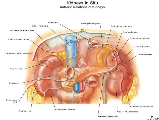
In brief, kidneys are a pair of organs that lie on either side of the vertebral column to be specific, they are in the posterior abdominal wall underneath the diaphragm. The key difference between left and right kidney is their position and size as the left kidney is slightly larger and higher in position than the right kidney.
Image Courtesy:
1. Gray1123 By Henry Vandyke Carter Henry Gray Anatomy of the Human Body Bartleby.com: Grays Anatomy, Plate 1123 via Commons Wikimedia
Also Check: Are Grapes Good For Kidney Stones