What Happens On The Day Of My Kidney Ultrasound
Unless told otherwise by your healthcare provider before the ultrasound, you can eat or drink as normal on the day of your test. If your provider needs a post void of your bladder, youll be required to drink 30 ounces of water an hour before the exam and not use the restroom until after the ultrasound.
Your ultrasound test will be performed by a registered, specially trained, technologist and interpreted by a board-certified radiologist.
Why Wait Until Your Kidneys Are Diseased
While the study was conducted on people with kidney disease, we could safely extrapolate the recommendations to those who want to avoid kidney disease and achieve optimal kidney function now, especially as we age.
In fact, additional research points to the actuality of physiological changes in the kidneys as we age. The research notes that a progressive reduction of the glomerular filtration rate and renal blood flow are observed in conjunction with aging. The reason for these phenomena is a decrease in the plasma flow in the glomerulus, a bundle of capillaries that partially form the renal corpuscle.2
In addition, the aging kidneys experience other structural changes, such as a loss of renal mass, and decreased responsiveness to stimuli that constrict or dilate blood vessels. The study concludes with a notable summation:
age-related changes in cardiovascular hemodynamics, such as reduced cardiac output and systemic hypertension, are likely to play a role in reducing renal perfusion and filtration. Finally, it is hypothesized that increases in cellular oxidative stress that accompany aging result in endothelial cell dysfunction and changes in vasoactive mediators resulting in increased atherosclerosis, hypertension and glomerulosclerosis.2
Signs You May Have Kidney Disease
More than 37 million American adults are living with kidney disease and most dont know it. There are a number of physical signs of kidney disease, but sometimes people attribute them to other conditions. Also, those with kidney disease tend not to experience symptoms until the very late stages, when the kidneys are failing or when there are large amounts of protein in the urine. This is one of the reasons why only 10% of people with chronic kidney disease know that they have it, says Dr. Joseph Vassalotti, Chief Medical Officer at the National Kidney Foundation.
While the only way to know for sure if you have kidney disease is to get tested, Dr. Vassalotti shares 10 possible signs you may have kidney disease. If youre at risk for kidney disease due to high blood pressure, diabetes, a family history of kidney failure or if youre older than age 60, its important to get tested annually for kidney disease. Be sure to mention any symptoms youre experiencing to your healthcare practitioner.
Don’t Miss: Liver Transplant Tattoo Ideas
Abdominal Ultrasound Scan: Tell Us Your Story
Have you or are you booked to have a scan of your abdomen? Any concerns, or perhaps did you get a scan report of your abdomen and wondering what it means? Post your abdominal ultrasound scan findings here and get help with interpretation of the jargons. Tell us your story or experience with an abdominal ultrasound scan.
Checking For Kidney Stones In The Emergency Department

First, the emergency doctor will give you medicine to help stop your pain. The medicine may be given by mouth. Or, it may be given through an intravenous needle placed in a vein in your arm. You may also be given medicine to help stop your nausea and vomiting. If you are dehydrated from vomiting, you may be given liquids through an IV tube.
Next, the emergency doctor will talk with you about your symptoms and medical history. If the emergency doctor thinks you might have a kidney stone, several tests may be done.
These may include:
- Urine Tests: To check for blood or mineral crystals in your urine or for signs of infection.
- Blood Tests: To check the health of your kidneys and for signs of a kidney or blood infection.
- Imaging Tests: To check for kidney stones in your urinary tract . Imaging tests may include a CT scan or an ultrasound.
Also Check: Can Apple Cider Vinegar Hurt Your Kidneys
Why Would I Need A Kidney And Bladder Ultrasound
Mayfair Mar 01, 2021
For bladder and kidney symptoms, such as pain, more or less frequent urination, uncomfortable urination, etc., its important to speak with your health care practitioner. Your doctor will likely order a number of tests to investigate the cause for these symptoms. These tests often include blood or urine tests, but medical imaging may also be recommended.
A kidney and bladder ultrasound, or renal ultrasound, uses high frequency sound waves to visualize and assess your kidneys, ureters and urinary bladder. For both men and women, this exam can help detect fluid collection, kidney or urinary tract infection, cysts, tumors, kidney disease, obstructions like kidney stones, and more.
While the severity of bladder and kidney conditions vary, many of them are very common. Its estimated between 40 to 60 percent of women develop a urinary tract infection during their lifetime, and the likelihood of an infection increases as you age. Kidney disease, on the other hand, affects one in 10 Canadians according to the Kidney Foundation of Canada.
Ultrasound imaging, also known as sonography, is often requested when investigating these bladder and kidney concerns, because its very good a looking at the soft tissues of the body, as well as evaluating blood flow and fluid retention.
What Happens During A Renal Scan
A renal scan is an outpatient, or same-day, procedure. You wont have to stay at the hospital overnight. A nuclear medicine technician performs the scan. This is usually done in either in a hospital radiology department or a medical office with special equipment.
Depending on the reasons for your scan, testing may take between 45 minutes and 3 hours. Talk to the technician beforehand if youre claustrophobic because the camera may pass close to your body.
Before your procedure, youll remove any of the following that could interfere with your scan:
- clothing
- dentures
- metal items
You may have to change into a hospital gown. Youll then lie down on a scanning table.
A technician may insert an intravenous line into a vein in your hand or arm. The technician will then insert a radioisotope into a vein in your arm. You may feel a quick, sharp poke with the injection.
There may be a waiting period between the injection and the first scan to allow your kidneys to process the radioisotope.
The scanner will detect the gamma rays from the radioisotope and create images of the area. Because any movement can alter or blur the image, youll need to stay still as the scanner creates an image.
If you need the scan because you have high blood pressure, you may receive a high blood pressure medication called an angiotensin converting enzyme inhibitor during testing. This allows for comparison of your kidneys before and after the medication is absorbed.
You May Like: Is Watermelon Good For The Kidneys
Ultrasound As A Nephrology Procedure
Sonography is essential to the practice of nephrology it is inexpensive and relatively easy to learn, making it an ideal tool to be incorporated into the practice of nephrology. With appropriate training, nephrologists can be competent at both performing and interpreting sonograms , and can provide the clinical correlation that is usually required for interpretation. However, nephrology has the dubious distinction of being one of the few specialties or subspecialties that has not embraced ultrasound and incorporated it into its training and practice. Although this void has been filled to some extent by the American Society of Diagnostic and Interventional Nephrology in the form of training guidelines and certification , few fellowship programs offer training that meets these guidelines . However, comprehensive training is available along with certification , and an increasing number of nephrology practices are incorporating this procedure. It is hoped that renal ultrasonography will eventually become an integral component of nephrology training.
Kidney Cysts Topic Overview
They could be associated with some disorders that eventually impair the function of the kidneys. But again more commonly, they are a type called simple kidney cysts that rarely cause serious complications. And this type is what were talking about in this section.
Causes
Its not fully known yet what causes these cysts. Currently, research suggests that they develop when the kidneys surface layer weakens and forms diverticulum which then fills with fluid, disengages, and develops into a cyst.
As noted before, age is often to blame the risk of having the condition increases with age. Furthermore, gender may also have an effect. Its relatively more common in men than in women.
Symptoms
The cyst looks like an oval or round fluid-filled pouch that typically has a well-defined outline. It usually develops in the surface of the kidney. However sometime it may also form inside the kidney.
*Image credit to Mayo
Most of the time, kidney cysts dont cause any symptoms. However, this doesnt mean that you can ignore them. Its still important to keep monitoring them!
How is kidney cyst diagnosed?
It is rarely to be concerned, and even it is often accidentally diagnosed. Many times it is discovered through an imaging test for another condition.
Standard procedures and tests to diagnose cysts in the kidneys include:
Complications
Don’t Miss: Can I Take Flomax Twice A Day For Kidney Stones
What Does A Healthy Kidney Look Like On An Ultrasound
Normal Kidneykidneykidneykidney
. Keeping this in consideration, what will a kidney ultrasound show?
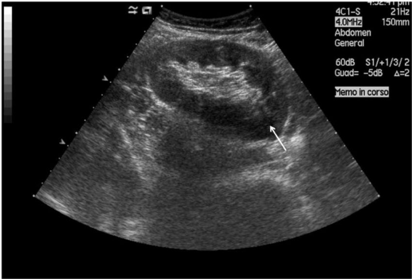
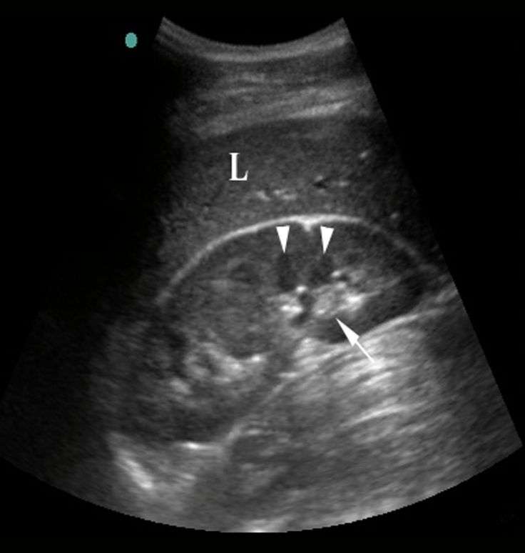
A kidney ultrasound may be used to assess the size, location, and shape of the kidneys and related structures, such as the ureters and bladder. Ultrasound can detect cysts, tumors, abscesses, obstructions, fluid collection, and infection within or around the kidneys.
Furthermore, can kidney disease be seen on ultrasound? Ultrasound scansIf you have been diagnosed with chronic kidney disease, you may be offered an ultrasound scan to help your doctor look for any problems with your kidneys. your blood tests show that your kidney disease is worsening you have blood in your urine
Accordingly, what do the colors mean on a kidney ultrasound?
The colors represent the speed and direction of blood flow within a certain area of the image . The mean velocity is then converted into a specific color. By definition, flow towards the transducer is depicted in red while flow away from the transducer is shown in blue.
Can you see kidney cancer in an ultrasound?
Most kidney cancers are found when people have an ultrasound or scan for an unrelated reason. The main tests for diagnosing kidney cancer are imaging scans and tissue sampling . Sometimes the doctor will also recommend an internal examination of the bladder, ureters and kidneys.
What Happens During My Exam
Ultrasound helps health care practitioners make a diagnosis and inform care decisions. Once your doctor has identified the need for an ultrasound, your doctors office may book an appointment for you, or provide you with a number to call to book your appointment. You will also be given a requisition form and preparation instructions for your exam.
Depending on the area to be examined, you may be asked to arrive with a full bladder or to fast and have nothing to eat or drink for six hours prior to your exam.
Once in the exam room you may be asked to change into a gown. You will then be positioned by one of our compassionate and experienced sonographers. A warm, unscented, hypo-allergenic ultrasound gel will be applied to the area of concern, and your sonographer will move the transducer around to gather images of your organs. You may be asked to hold your breath and change position to help better examine the area of concern. You may experience mild to moderate pressure while the sonographer takes the images.
Don’t Miss: How Does Flomax Help With Kidney Stones
Autosomal Dominant Polycystic Kidneys With Multiple Calculi
this 58-year-old Male patient underwent routine sonography for non-specific complaints including mildly elevated serum creatinine and blood urea. Sonography shows multiple small cysts ranging from3 mm up to 12 mm in size in both kidneys. Both kidneys are literally studded with these minute cystic lesions with many of them showing the presence of milk of calcium.In addition, there are also multiple minute calculi ranging from 2 to 4 mm in size. This is an unusual presentation of autosomal dominant polycystic kidney disease and could represent the initial stages of the disease. Both kidneys also show moderate enlargement with sizes of 11 x 6 cm.
Procedural Applications Of Ultrasound In Nephrology

Because of its ease of use and availability at the bedside, sonography has revolutionized the performance of percutaneous procedures. The enhanced success rates and reduced complication rates with ultrasound guidance have made it the standard of care for many of these procedures, including those procedures performed by nephrologists. However, like any tool, success depends on proper use and an understanding of the limitations. There are two basic approaches. The entry site, angle, and depth can be determined with ultrasound, after which the needle is placed without direct ultrasound guidance , or ultrasound can be used during the needle insertion .
Also Check: Celery Juice Kidneys
What To Expect After A Kidney Ultrasound
Youll be able to eat and drink as usual after your ultrasound. Additionally, you can return to your daily activities after you leave the facility.
After the ultrasound, the technician will forward the results to a radiologist. This is a type of doctor that specializes in making sense of medical images, such as those created by ultrasound.
Once the radiologist has reviewed your images, which typically takes only 1 or 2 days, theyll send their findings to your doctor. Your doctor will then contact you to go over the results of your ultrasound.
When Is A Renal Ultrasound Recommended
When blood or urine tests expose the presence of abnormalities in the renal system, your doctor may recommend a renal ultrasound. In addition to detecting cysts, tumors and stones, ultrasound can:
- Reveal harmful abscesses, fluid collection, infection within or around the kidneys
- Help ensure accurate needle placement for biopsy
- Place a drainage tube
Don’t Miss: Is Red Wine Good For Kidney Stones
How You Have An Ultrasound Scan
The ultrasound scanner has a microphone that gives off sound waves. The sound waves bounce off the organs inside your body, and the microphone picks them up. The microphone links to a computer that turns the sound waves into a picture on the screen.
Ultrasound scans are completely painless. You usually have the scan in the hospital x-ray department by a sonographer. A sonographer is a trained professional who is specialised in ultrasound scanning.
You might need to change position to lie on your side or your front for a few minutes.
I Introduction And Indications
- Point-of-care renal ultrasound is a rapid, bedside test for the evaluation of the patient with suspected renal colic or urinary retention.
- Because ureteral stones can be difficult to visualize by US,1 the secondary finding of hydronephrosis is used to diagnose nephrolithiasis when the clinical suspicion for renal colic is high.
- POC renal US for the diagnosis of nephrolithiasis has a reported sensitivity and specificity of 70% and 75%, respectively using the gold standard of CT examination2 and can decrease cumulative radiation exposure.3
- POC US should be used as part of a treatment pathway to safely diagnose and disposition patients with uncomplicated renal colic. Studies demonstrate that patients with mild hydronephrosis on POC US are less likely to have large ureteral calculi or require urologic intervention4-6 and those with moderate to severe hydronephrosis are likely to have stones > 5mm in size.6,7
- POC US can also be used to estimate bladder volume for the diagnose of urinary retention.8-11
Read Also: Soda Cause Kidney Stones
How Do The Kidneys Work
The body takes nutrients from food and converts them to energy. Afterthe body has taken the food that it needs, waste products are leftbehind in the bowel and in the blood.
The kidneys and urinary system keep chemicals, such as potassium andsodium, and water in balance, and remove a type of waste, called urea,from the blood. Urea is produced when foods containing protein, such asmeat, poultry, and certain vegetables, are broken down in the body.Urea is carried in the bloodstream to the kidneys.
Two kidneys, a pair of purplish-brown organs, are located below theribs toward the middle of the back. Their function is to:
-
Remove liquid waste from the blood in the form of urine
-
Keep a stable balance of salts and other substances in the blood
-
Produce erythropoietin, a hormone that aids the formation of red blood cells
-
Regulate blood pressure
The kidneys remove urea from the blood through tiny filtering unitscalled nephrons. Each nephron consists of a ball formed of small bloodcapillaries, called a glomerulus, and a small tube called a renaltubule.
Urea, together with water and other waste substances, forms the urineas it passes through the nephrons and down the renal tubules of thekidney.
What Are The Reasons For A Kidney Ultrasound
A kidney ultrasound may be used to assess the size, location, and shapeof the kidneys and related structures, such as the ureters and bladder.Ultrasound can detect cysts, tumors, abscesses, obstructions, fluidcollection, and infection within or around the kidneys.Calculi of the kidneysand ureters may be detected by ultrasound.
A kidney ultrasound may be performed to assist in placement of needlesused tobiopsy the kidneys, to drain fluid from a cyst or abscess, or to place a drainage tube.This procedure may also be used to determine blood flow to the kidneysthrough the renal arteries and veins.
Kidney ultrasound may be used after akidney transplantto evaluate the transplanted kidney.
There may be other reasons for your physician to recommend a kidneyultrasound.
Our Approach to Kidney Ultrasounds
The Johns Hopkins Kidney Program is one of the first in the country and our doctors have pioneered some of the most innovative treatments for patients with renal failure. In addition to offering leading-edge procedures, our program offers shorter wait times and minimally invasive options, which can lead to a faster recovery.
Also Check: Pineapple Juice And Kidney Stones