Electrical Impulses Keep The Beat
The heart’s four chambers pump in an organized manner with the help of electrical impulses that originate in the sinoatrial node . Situated on the wall of the right atrium, this small cluster of specialized cells is the heart’s natural pacemaker, initiating electrical impulses at a normal rate.
The impulse spreads through the walls of the right and left atria, causing them to contract, forcing blood into the ventricles. The impulse then reaches the atrioventricular node, which acts as an electrical bridge for impulses to travel from the atria to the ventricles. From there, a pathway of fibers carries the impulse into the ventricles, which contract and force blood out of the heart.
Which Blood Vessels Delivers Blood To The Cortex
The brain receives blood from two sources: the internal carotid arteries, which arise at the point in the neck where the common carotid arteries bifurcate, and the vertebral arteries . The internal carotid arteries branch to form two major cerebral arteries, the anterior and middle cerebral arteries.
What Is The Function Of The Interlobular Artery
4.3/5arteriesinterlobular arteriesarteries
Renal artery carries mineral rich, oxygenated blood from the heart to the kidneys for nutrition and cellular respiration. Renal veins carry deoxygenated blood after waste products have been removed via glomerular filtration back from the kidneys to the heart.
Likewise, what does the renal artery divide into? Renal CirculationEach renal artery arises directly from the abdominal aorta and once it passes through the hilum, divides into a number of interlobar arteries, which supply each lobule of the kidney.
Also asked, where is the Interlobar artery?
The Renal Arteries Arise from the Abdominal AortaThe single renal artery enters the hilum and then branches to form the interlobar arteries, so-named because they pass between the lobes of the kidney. At the junction of the cortex and medulla, the interlobar arteries bend over to form incomplete arches.
What artery delivers blood to the glomerulus?
afferent arteriole
Read Also: Is Grape Juice Good For Kidneys
Renal Artery Stenosis: Symptoms & Signs
- Medical Author: Melissa Conrad Stöppler, MD
Medically Reviewed on 7/1/2020
Renal artery stenosis refers to a narrowing of the blood vessel that delivers blood to the kidney for filtration . This leads to a restriction of blood flow to the kidney, and particularly when the arteries to both kidneys are affected, may lead to impaired kidney function and high blood pressure .
Renal artery stenosis typically does not cause specific signs or associated symptoms. Over time, it may cause problems related to declining kidney function such as
- elevated protein levels in the urine or
- other signs of abnormal kidney function.
Causes of renal artery stenosis
Most commonly, atherosclerosis causes renal artery stenosis. High cholesterol levels, high blood pressure, smoking, and diabetes all increase the risk of atherosclerosis.
Other renal artery stenosis symptoms and signs
- Abnormal Kidney Function
The Superior Vena Cava

The superior vena cava drains most of the body superior to the diaphragm. On both the left and right sides, the subclavian vein forms when the axillary vein passes through the body wall from the axillary region. It fuses with the external and internal jugular veins from the head and neck to form the brachiocephalic vein. Each vertebral vein also flows into the brachiocephalic vein close to this fusion. These veins arise from the base of the brain and the cervical region of the spinal cord, and flow largely through the intervertebral foramina in the cervical vertebrae. They are the counterparts of the vertebral arteries. Each internal thoracic vein, also known as an internal mammary vein, drains the anterior surface of the chest wall and flows into the brachiocephalic vein.
The azygos vein passes through the diaphragm from the thoracic cavity on the right side of the vertebral column and begins in the lumbar region of the thoracic cavity. It flows into the superior vena cava at approximately the level of T2, making a significant contribution to the flow of blood. It combines with the two large left and right brachiocephalic veins to form the superior vena cava.
Figure 14 and Table 9 summarize the veins of the thoracic region that flow into the superior vena cava.
Figure 14. Veins of the thoracic and abdominal regions drain blood from the area above the diaphragm, returning it to the right atrium via the superior vena cava.
You May Like: Is Pineapple Good For Kidney Stones
Blood Vessels Of The Kidney
The renal arteries are both branches of the abdominal aorta. The right renal artery passes dorsal to the posterior vena cava. On entering the kidney at the hilum, the renal arteries divide into three or four branches that run dorsally and ventrally around the pelvis of the kidney to reach the junction between the cortex and subcortex, where they form the arcuate arteries.
Interlobar arteries are end arteries that have no anastomotic connections with other arteries, either in the rat or in man. The arcuate arteries run at the junction of the cortex and the outer stripe of the outer medulla where they give rise to the interlobular arteries that run at right angles from the arcuate arteries towards the surface of the cortex.
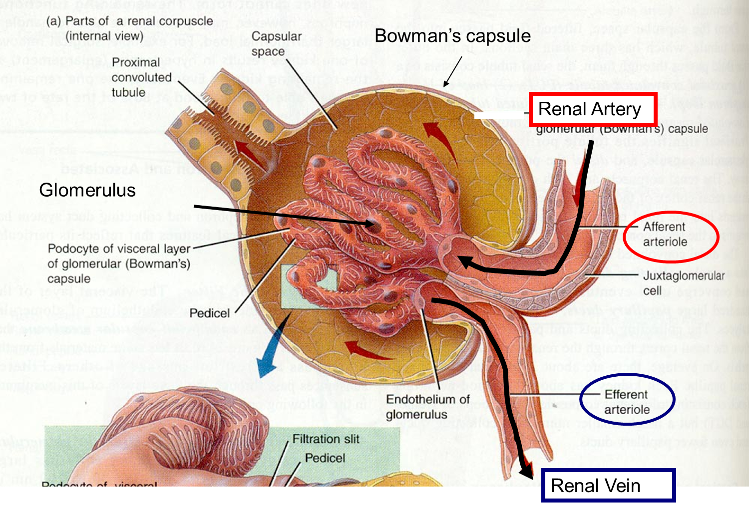
The interlobular arteries give rise to the afferent arterioles of the glomeruli that lie within the Bowmans capsules of the Malpighian corpuscles, and then recombine to produce the efferent arterioles. Afferent arterioles leaving interlobular arteries deep in the cortex point downwards towards the medulla; those leaving nearer the surface of the kidney point towards the surface.
E.J. Johns, A.F. Ahmeda, in, 2014
What Surgical Procedures Are Available For Renal Artery Stenosis
If the results of any of these screening tests suggest an abnormality of the renal artery, an x-ray angiography is then performed. A 75% or greater narrowing of the renal artery seen on the angiogram has been termed treatable renal artery stenosis.
Treatable means that the stenosis of the artery is severe , the artery needs to be widened , and it has a good chance of responding favorably to the dilatation. Usually right at the time of the angiography, an angioplasty is done. In this procedure a tiny balloon is inflated in the interior space in the artery to dilate the narrowed artery. Additionally, as part of the angioplasty procedure, a stent may be placed in the artery.
In rare cases, vascular surgery may be done for renal artery stenosis. In these situations, typically another vascular surgery near the renal arteries, for example the aorta, is the main procedure. If renal artery stenosis is also present, then a bypass renal artery surgery may be done at the same time.
These invasive procedures are typically reserved for cases that do not respond to medical treatment and where it has been determined that the stenosis is causing or contributing to the uncontrolled high blood pressure. These invasive procedures may only be done if it is thought that the kidney dysfunction or elevated blood pressure can be effectively treated with the procedures.
You May Like: Does Red Wine Cause Kidney Stones
Facts You Should Know About Renal Artery Stenosis
- Elevated blood pressure is common and is generally simply treated with medications.
- Likewise, various other methods are used to treat the large majority of patients with kidney failure.
- There is a small subgroup of patients with high blood pressure;and/or renal failure caused by renal artery stenosis.
- Some of these patients may respond favorably to dilating the narrowed artery, using the technique of angioplasty.
- The patients that can benefit from angioplasty have a severe stenosis of the renal artery and do not have a very high renal vascular resistance.
Veins Of The Head And Neck
Blood from the brain and the superficial facial vein flow into each internal jugular vein. Blood from the more superficial portions of the head, scalp, and cranial regions, including the temporal vein and maxillary vein, flow into each external jugular vein. Although the external and internal jugular veins are separate vessels, there are anastomoses between them close to the thoracic region. Blood from the external jugular vein empties into the subclavian vein. Table 10;summarizes the major veins of the head and neck.
| Table 10. Major Veins of the Head and Neck | |
|---|---|
| Vessel | |
| Drains blood from the maxillary region and flows into the external jugular vein | |
| External jugular vein | Drains blood from the more superficial portions of the head, scalp, and cranial regions, and leads to the subclavian vein |
Also Check: Is Mulberry Good For Kidneys
Causes Of Renal Vascular Disease
The most common cause of renal artery blockages is arteriosclerosis with cholesterol and plaque build-up. This is similar to what is seen in the coronary arteries of the heart, the carotid arteries to the brain and the leg vessels. Men are affected with this condition twice as often as women, with 55 the average age at diagnosis. Other causes of renal vascular disease include:
- Fibromuscular dysplasia , an abnormal growth on the inside of the renal artery that typically affects middle-aged women, but has been found in men and people of all ages.
- Renal artery aneurysms can often twist or compress a nearby renal artery, causing it to become narrowed.
Overview Of Systemic Veins
Systemic veins return blood to the right atrium. Since the blood has already passed through the systemic capillaries, it will be relatively low in oxygen concentration. In many cases, there will be veins draining organs and regions of the body with the same name as the arteries that supplied these regions and the two often parallel one another. This is often described as a complementary pattern. However, there is a great deal more variability in the venous circulation than normally occurs in the arteries. For the sake of brevity and clarity, this text will discuss only the most commonly encountered patterns. However, keep this variation in mind when you move from the classroom to clinical practice.
In both the neck and limb regions, there are often both superficial and deeper levels of veins. The deeper veins generally correspond to the complementary arteries. The superficial veins do not normally have direct arterial counterparts, but in addition to returning blood, they also make contributions to the maintenance of body temperature. When the ambient temperature is warm, more blood is diverted to the superficial veins where heat can be more easily dissipated to the environment. In colder weather, there is more constriction of the superficial veins and blood is diverted deeper where the body can retain more of the heat.
Figure 13. The major systemic veins of the body are shown here in an anterior view.
Recommended Reading: Can Kidney Stones Affect Your Psa Count
What Are The Effects Of Vascular Disease
Because the functions of the blood vessels include supplying all organs and tissues of the body with oxygen and nutrients, removal of waste products, fluid balance, and other functions, conditions that affect the vascular system may affect the part of the body supplied by a particular vascular network, such as the coronary arteries of the heart.
Examples of the effects of vascular disease include:
-
Coronary artery disease. Heart attack, angina
-
Cerebrovascular disease. Stroke, transient ischemic attack
-
Peripheral arterial disease. Claudication , critical limb ischemia
-
Vascular disease of the great vessels. Aortic aneurysm , coarctation of the aorta , Takayasu arteritis
-
Thoracic vascular disease. Thoracic aortic aneurysm
-
Abdominal vascular disease. Abdominal aortic aneurysm
-
Peripheral venous disease. Deep vein thrombosis , varicose veins
-
Lymphatic vascular diseases. Lymphedema
-
Vascular diseases of the lungs. Granulomatosis with polyangiitis , angiitis , hypertensive pulmonary vascular disease
-
Renal vascular diseases. Renal artery stenosis , fibromuscular dysplasia
-
Genitourinary vascular diseases. Vascular erectile dysfunction
Vasculature And Innervation Of The Kidney

The kidney receives 25% of the cardiac output, with the cortex receiving 850% of renal blood flow. The outer medulla receives 14% and the inner medulla 1%. In the adult kidney, the renal artery branches in the pelvis into 6 to 10 interlobar arteries, giving rise in turn to arcuate arteries coursing parallel to the capsule along the corticomedullary junction. Interlobular arteries arise from the arcuates, coursing perpendicular to the capsule, each supplying a cortical labyrinth. Each interlobular artery gives rise to 6 to 11 afferent arterioles. All renal artery branches are end arteries without significant collateral supply. The renal circulation is a portal system as capillary beds reunite into an efferent arteriole, running for a varying distance before again forming a capillary bed surrounding portions of the nephron. All circulation to the medulla is of postglomerular capillary derivation. Veins run parallel to the main arterial and arcuate system. Lymphatics run parallel only to the cortical vasculature, being absent in the medulla. The interstitium of cortex and medulla are not functionally contiguous.
Nerves that are associated with intrarenal arterioles have been noted to have ramifications in the afferent and efferent arterioles, as well as in the juxtaglomerular apparatus. These nerve fibers are monoaminergic with norepinephrine and dopamine activity.
Peter Greaves MBChB FRCPath, in, 2012
Also Check: Is Grape Juice Good For Kidney Stones
The Inferior Vena Cava
Other than the small amount of blood drained by the azygos and hemiazygos veins, most of the blood inferior to the diaphragm drains into the inferior vena cava before it is returned to the heart . Lying just beneath the parietal peritoneum in the abdominal cavity, the inferior vena cava parallels the abdominal aorta, where it can receive blood from abdominal veins. The lumbar portions of the abdominal wall and spinal cord are drained by a series of lumbar veins, usually four on each side. The ascending lumbar veins drain into either the azygos vein on the right or the hemiazygos vein on the left, and return to the superior vena cava. The remaining lumbar veins drain directly into the inferior vena cava.
Blood supply from the kidneys flows into each renal vein, normally the largest veins entering the inferior vena cava. A number of other, smaller veins empty into the left renal vein. Each adrenal vein drains the adrenal or suprarenal glands located immediately superior to the kidneys. The right adrenal vein enters the inferior vena cava directly, whereas the left adrenal vein enters the left renal vein.
From the male reproductive organs, each testicular vein flows from the scrotum, forming a portion of the spermatic cord. Each ovarian vein drains an ovary in females. Each of these veins is generically called a gonadal vein. The right gonadal vein empties directly into the inferior vena cava, and the left gonadal vein empties into the left renal vein.
What Organs Are Attached To The Kidneys
5/5ureterbladderfull answer
Your kidneys are shaped like beans, and each is about the size of a fist. They are near the middle of your back, one on either side of your spine, just below your rib cage. Each kidney is connected to your bladder by a thin tube called a ureter.
Furthermore, which part of the body the kidney is located? Kidney pain definition and factsThe function and purpose of the kidneys are to remove excess fluid and waste products from the body. The kidneys are organs that are located in the upper abdominal area against the back muscles on both the left and right side of the body.
Keeping this in view, what organ does the renal artery supply?
The renal arteries normally arise off the left interior side of the abdominal aorta, immediately below the superior mesenteric artery, and supply the kidneys with blood. Each is directed across the crus of the diaphragm, so as to form nearly a right angle.
What organs are above the kidneys?
Adrenal Disease. Each person is usually born with two adrenal glands. The adrenals are paired, goldenrod-yellow colored glands that are situated behind the organs of the gastrointestinal tract, next to the spine, and just above the kidneys, in a space called the retroperitoneum.
Some conditions that cause loss of blood flow to the kidneys include:
- a heart attack.
- scarring of the liver or liver failure.
- dehydration.
Don’t Miss: Does Red Wine Cause Kidney Stones
How Common Is Renal Artery Stenosis
Narrowing of the kidney arteries is more common in individuals 50 years of age and older. It is estimated that some degree of narrowing is found in about 18% of adults between 65-75 years of age and 42% of those older than 75 years of age. This may be due to the fact that atherosclerosis is more common in this age group.
In younger patients, the narrowing of the renal artery usually is due to the thickening of the artery and it is more common in women than men.
It is estimated that renal artery stenosis accounts for approximately 1% of mild to moderate cases of high blood pressure. It may be responsible for more than 10% of cases of severely elevated or difficult to treat high blood pressure .
What Are The Renal Arteries
Renal refers to anything related to the kidneys. Renal arteries carry blood from the heart to the kidneys. They branch directly from the aorta on either side and extend to each kidney. These arteries take a very large volume of blood to the kidneys to be filtered.
The heart pumps out approximately 5 liters of blood per minute, and about 1-1.5 liters of the total volume of blood pumped by the heart passes through the kidneys every minute.
Recommended Reading: Is Pineapple Good For Kidney Stones
What Is Arcuate Artery
4.9/5arcuate arteryarteriesarteryarteriesarcuate arteries
The arcuate artery supplies blood to the adjoining muscles, metatarsal bones and toes.
Additionally, what are the arteries for? The arteries are the blood vessels that deliver oxygen-rich blood from the heart to the tissues of the body. Each artery is a muscular tube lined by smooth tissue and has three layers: The intima, the inner layer lined by a smooth tissue called endothelium.
Also to know is, what is the function of the arcuate vein?
Arcuate Veins:These veins receive oxygen poor blood from the renal cortical veins and drain it into the interlobar veins , before it exits via the renal vein .
Is there a major artery in your foot?
Posterior tibial artery: This branch of the popliteal artery supplies oxygenated blood to the leg and sole of the foot. Plantar arteries: The plantar arterieslateral, medial, and deepform a looping web of arteries across the foot and down through each toe. They eventually unite with the dorsalis pedis artery.