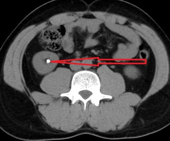
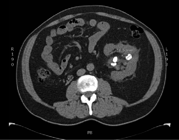
Ultrasound For Kidney Stones
Your doctor might recommend an ultrasound for kidney stones because it is a quick, safe and easy procedure. An ultrasound uses sound waves to create images and does not involve radiation.
During an ultrasound, youll lie on an exam table while a technologist moves a transducer over the part of the body being scanned. A transducer is a handheld device that sends and receives sound waves. The sound waves will then be processed by a computer to produce images.
An ultrasound may provide enough evidence for a kidney stone diagnosis. However, if the images are not clear, your doctor might order a computer tomography scan.
What Are The Reasons For A Ct Scan Of The Kidney
A CT scan of the kidney may be performed to assess the kidneys fortumors and other lesions, obstructions such askidney stones, abscesses,polycystic kidney disease, and congenital anomalies, particularly when another type ofexamination, such as X-rays or physical examination, is not conclusive.CT scans of the kidney may be used to evaluate the retroperitoneum . CT scansof the kidney may be used to assist in needle placement inkidney biopsies.
After the removal of a kidney, CT scans may be used to locate abnormalmasses in the empty space where the kidney once was. CT scans of thekidneys may be performed afterkidney transplantsto evaluate the size and location of the new kidney in relation to thebladder.
There may be other reasons for your doctor to recommend a CT scan ofthe kidney.
What Happens During A Ct Scan
CT scans may be performed on an outpatient basis or as part of yourstay in a hospital. Procedures may vary depending on your condition andyour physician’s practices.
Generally, a CT scan follows this process:
You may be asked to change into a patient gown. If so, a gown will be provided for you. A locked will be provided to secure all personal belongings. Please remove all piercings and leave all jewelry and valuables at home.
If you are to have a procedure done with contrast, an intravenous line will be started in the hand or arm for injection of the contrast media. For oral contrast, you will be given a liquid contrast preparation to swallow. In some situations, the contrast may be given rectally.
You will lie on a scan table that slides into a large, circular opening of the scanning machine. Pillows and straps may be used to prevent movement during the procedure.
The technologist will be in another room where the scanner controls are located. However, you will be in constant sight of the technologist through a window. Speakers inside the scanner will enable the technologist to communicate with and hear you. You may have a call button so that you can let the technologist know if you have any problems during the procedure. The technologist will be watching you at all times and will be in constant communication.
As the scanner begins to rotate around you, X-rays will pass through the body for short amounts of time. You will hear clicking sounds, which are normal.
Read Also: Does Red Wine Cause Kidney Stones
How Do The Kidneys Work
The body takes nutrients from food and converts them to energy. Afterthe body has taken the food that it needs, waste products are leftbehind in the bowel and in the blood.
The kidneys and urinary system keep chemicals, such as potassium andsodium, and water in balance, and remove a type of waste, called urea,from the blood. Urea is produced when foods containing protein, such asmeat, poultry, and certain vegetables, are broken down in the body.Urea is carried in the bloodstream to the kidneys.
Two kidneys, a pair of purplish-brown organs, are located below theribs toward the middle of the back. Their function is to:
-
Remove liquid waste from the blood in the form of urine
-
Keep a stable balance of salts and other substances in the blood
-
Produce erythropoietin, a hormone that aids the formation of red blood cells.
-
Regulate blood pressure
The kidneys remove urea from the blood through tiny filtering unitscalled nephrons. Each nephron consists of a ball formed of small bloodcapillaries, called a glomerulus, and a small tube called a renaltubule.
Urea, together with water and other waste substances, forms the urineas it passes through the nephrons and down the renal tubules of thekidney.
When Urinalysis And Other Urine Tests Help In Kidney Stone Diagnosis

In addition to imaging tests, doctors usually order urine tests to help determine what type of stone you may have and why you are developing stones. This information can help your doctor better advise you about how to prevent future kidney stones, says Naim Maalouf, MD, an associate professor of internal medicine at UT Southwestern Medical Center in Dallas. At least 31 percent of people diagnosed with kidney stones develop another one within 10 years.
Kidney stones are made of minerals and other substances that can be found in the urine that can be identified with testing. Types of kidney stones include calcium stones , uric acid stones, struvite stones, and cystine stones.
Notably, a urinalysis test and urine culture can also tell doctors whether you also have an infection, which is a potentially life-threatening complication in combination with a kidney stone, says Seth K. Bechis, MD, an assistant professor of urology at UC San Diego Health in California. If urine is trapped behind an obstructing stone in the ureter, urine can become infected. This scenario can cause an infection of the kidney tissue or spread to the bloodstream.
Doctors may perform the following urine tests in kidney stone diagnosis:
A urinalysis with microscopy can also help doctors find evidence of bleeding or infection, says Dr. Maalouf.
From this urine sample, doctors can tell whether people are predisposed to stone formation. We call it a stone risk profile, says Maalouf.
Recommended Reading: Grapes And Kidney Stones
Ultrasound Or Ct In Diagnosis Of Kidney Stones
Two tests are available for diagnosing kidney stones, CT scan or renal ultrasound. With wide availability of CT scans the pendulum has swung towards using CT scans for diagnosis of kidney stones.
CT scan is easy to perform. While a single CT scan in adults is fairly safe and is associated with fairly small amount of radiation, many patients with kidney stones will have multiple recurrent episodes of kidney stones requiring multiple CT scans during their life. I have seen patients who had 5-10 CAT scans in a period of 1-2 years for evaluation of kidney stones. These patients have been exposed to significant amount of radiation in a very short period of time which is very concerning.
In addition they are likely to have a need for CT scan for evaluation of other symptoms at some point in their life. There is significant evidence that accumulated radiation from exposure to multiple CT scans can have detrimental effect on life and may be associated with a small risk of developing certain cancers. Radiation induced cancers typically do not develop immediately but take many years to occur.
While sometimes CT scan provides valuable information that cannot be obtained with other methods, in my practice over the past 5 years the use of CT scans for evaluation and management of patients with kidney stones has decreased while the use of ultrasound has increased.
Information Gained From Imaging
The emergency department is a common setting for the initial presentation of patients with obstructing stones. Such a diagnosis might be suspected without imaging however practitioners must entertain a wide differential diagnosis for patients with severe abdominal and/or flank pain. Imaging modalities with high sensitivity provide the clinician with confidence that symptoms are caused by an alternative pathology when no stones are visualized. Alternatively, imaging modalities with high specificity demonstrate that a patientâs symptoms are related to stones when they are visualized. Measurements of sensitivity and specificity can vary widely throughout the literature based on several factors, including the method used as the reference standard to determine true-positive and true-negative values, and the population of patients being examined.
Broadly, the available imaging modalities include CT, ultrasonography, KUB radiography, and MRI. The sensitivity, specificity, dose of ionizing radiation, and relative costs vary between modalities . An algorithm is also proposed for imaging patients with suspected stones in the emergency department setting .
A proposed algorithm for imaging patients with acute stone disease in the emergency department
Recommended Reading: Is Cranberry Juice Good For Your Liver And Kidneys
Ultrasound As Good As Ct For Initial Diagnosis Of Kidney Stones : Study
By Gene Emery, Reuters Health
6 Min Read
NEW YORK – Using the sound waves of an ultrasound to detect a painful kidney stone is just as effective as the X-rays of a CT scan, and exposes patients to much less harmful radiation, according to a new multicenter study.
Its actually quite surprising that ultrasound is just as good as CT scanning when you look at patient outcomes, said Dr. Rebecca Smith-Bindman of the University of California, San Francisco, chief author of the report in the New England Journal of Medicine.
Dr. Charles D. Scales, Jr. of Duke University Medical Center in Durham, North Carolina, called it a really provocative study adding, it should make doctors and patients think about what we do when a kidney stone may be causing a patients pain.
It doesnt necessarily say patients should not get a CT scan, said Scales, who was not connected with the research, but I think the main message is that an ultrasound is the best place to start.
Kidney stones account for nearly a million emergency room visits in the U.S. each year at a cost of nearly a billion dollars. One in 11 Americans say they have had one.
For years, an abdominal CT scan has been the standard method for detecting stones because it makes the stones easier to see than regular X-rays. But other calcium deposits in the body can be mistaken for stones, leading to unnecessary treatment.
SOURCE: bit.ly/1ARC2GG New England Journal of Medicine, September 17, 2014.
Diagnosis Of Kidney Stones
Your doctor will ask you questions about your medical history, what medications you are currently taking, and whether anyone in your family suffers from kidney stones.
Your doctor may take a sample of your urine to see if there is an infection.
Anx-ray shows certain types of kidney stones. Most kidney stones can be seen on an x-ray. This test is helpful for knowing what type of stone you may have. Other studies are often needed to determine the specific spot in the kidney where the stone is located.
For this test, a dye is injected into a vein. The dye highlights otherwise hard-to-see areas of your urinary tract as it passes out of your system. This makes it easier for your doctor to see the kidney stone on an X-ray.This procedure is less commonly used today because of the excellent images obtained with CT scans.
This procedure uses X-rays to take highly detailed pictures of your internal organs. A CT scan can spot small kidney stones that regular X-rays might miss.
Blood tests help identify factors such as high levels of calcium, uric acid, or the presence of infection that can cause kidney stones to develop.
Urine will be tested for acidity and levels of substances, like calcium, uric acid, citrate, and oxalate, that can form kidney stones. This test provides a more accurate analysis than your doctor would get from a single urine sample. This sample is collected over a 24-hour period.
Stone Analysis
Read Also: Cranberry Good For Liver
How Are Kidney And Bladder Stones Diagnosed And Evaluated
Imaging is used to provide your doctor with valuable information about the kidney or bladder stones, such as location, size and effect on the function of the kidneys. Some types of imaging that your doctor may order include:
- Abdominal and pelvic CT: This is the most rapid scanning method for locating a stone. This procedure can provide detailed images of the kidneys, ureters, bladder and urethra, identify a stone and reveal whether it is blocking urinary flow. See the Safety page for more information about CT procedures.
- Intravenous pyelogram : This is an x-ray examination of the kidneys, ureters and urinary bladder that uses iodinated contrast material injected into veins to evaluate the urinary system. See the Safety page for more information about x-rays.
- Abdominal and Pelvic ultrasound: These exams use sound waves to provide pictures of the kidneys and bladder and can identify blockage of urinary flow and help identify stones.
For more information about ultrasound performed on children, visit the pediatric abdominal ultrasound page.
Diagnostic Tests For Kidney Stones
Your doctor conducts an initial physical exam and may order several tests, including blood and urine tests and imaging exams, to determine whether you have kidney stones and to diagnose the particular type and location. If you have passed a stone or had one surgically treated, your doctor may analyze the stone to determine its type and suggest additional testing to find out if you have more.
All of these tests may be scheduled on the same day as your doctors visit. Our radiologists and urologists can also review the results of scans youve had done at other medical facilities.
Read Also: Does Carbonated Water Cause Kidney Stones
Rising Rates Of Kidney Stones
Kidney stone rates are increasing, and in a 2010 National Health and Nutrition Examination Survey, one in 11 people reported having had at least one kidney stone. The use of CT to diagnose kidney stones has risen 10-fold in the last 15 years. CT exams generally are conducted by radiologists, while ultrasound exams may be conducted by emergency room physicians as well as radiologists.
An ultrasound image of the bladder showing a ureter blocked by a kidney stone.
Emergency room patients whose pain was suspected to be due to kidney stones were randomly assigned in the NEJM study to one of three imaging groups. In one group patients received an ultrasound exam performed by an emergency room physician on site. A second group received similar ultrasonography performed by a radiologist, a specialist in the procedure. The third group received an abdominal CT scan, also conducted by a radiologist.
With six months of patient follow-up, the study found that health outcomes for 2,759 patients were just as good with ultrasound as with CT, and that patients fared no worse when emergency physicians instead of radiologists performed the ultrasound exam. Serious adverse events, pain, return trips to the emergency department or hospitalizations did not differ significantly among groups.
The study sites were emergency departments at academic medical centers throughout the country, and included four safety-net hospitals serving low-income communities.
More Information At No Additional Cost

If a CT scan is ordered, we should put it to good use. Opportunistic CT imaging does not add cost or radiation exposure, nor does it require additional equipment. It can identify potentially significant comorbidities that may require further evaluation and management . It can predict underlying metabolic abnormalities, suggest kidney stone preventive strategies and identify those best suited for a trial of dissolution therapy.
The prevalence of nephrolithiasis in the United States is approximately 9 percent and the disease carries significant burden to patients and the health care system. Our future studies will evaluate the impact of medical approaches to stone prevention on bone mineral density. The goal is to maximize the opportunity our imaging provides.
Dr. Patel, a former fellow, is now an assistant professor at UCLA Health in Los Angeles. Dr. Ward is a diagnostic radiology resident.
Read Also: Can Seltzer Water Cause Kidney Stones
Will Kidney Stones Show Up On Ultrasound
Ask U.S. doctors your own question and get educational, text answers â it’s anonymous and free!
Ask U.S. doctors your own question and get educational, text answers â it’s anonymous and free!
HealthTap doctors are based in the U.S., board certified, and available by text or video.
Things To Discuss With A Doctor
If a person is experiencing any urinary tract symptoms, they should contact a doctor for advice.
Without treatment, urinary tract symptoms can cause severe health issues. Likewise, untreated kidney infections can lead to problems such as kidney disease, high blood pressure, or kidney failure.
UTIs during pregnancy can also be dangerous for both the pregnant person and the fetus.
If kidney stones remain untreated, they can block the urinary tubes, which increases the risk of infection and can put a strain on the kidneys.
If a doctor recommends a CT urogram and the person has any concerns about the procedure, they should discuss the possibility of having a mild sedative. Mild sedation can help alleviate anxiety and any feelings of claustrophobia during the procedure.
Recommended Reading: Are Almonds Bad For Your Kidneys
What Happens During A Ct Scan Of The Kidney
You may have a CT scan as an outpatient or as part of your stay in a hospital.
The way the test is done may vary depending on your condition and your healthcare provider’s practices.
Generally, a CT scan of the kidney follows this process: