Structural And Functional Inter
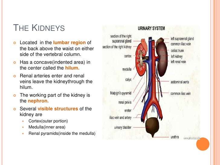
An adult typically has two kidneys, which are reddish-brown and bean-shaped . Although their size can vary, the length of the average kidney is about 11 cm, the thickness about 2.5 cm, and the width is about 5 cm. The average weight of a single kidney is 15 g. The kidney’s lateral border is convex, whereas the medial border is concave and indented in a depression called the renal hilus. All related structures enter or leave the kidney at the hilus .
The kidneys lie in the retroperitoneal space of the posterior abdominal cavity. Thus, they can be exposed without opening the peritoneal cavity. One kidney lies on either side of the vertebral column. Layers of muscle surround the posterior surfaces of the kidneys, and abdominal organs surround their anterior surfaces. The peritoneal membrane covers most of the anterior surface of each kidney.
| The renal capsule and the renal fascia |
Where Are The Renal Pyramids Located In The Kidney
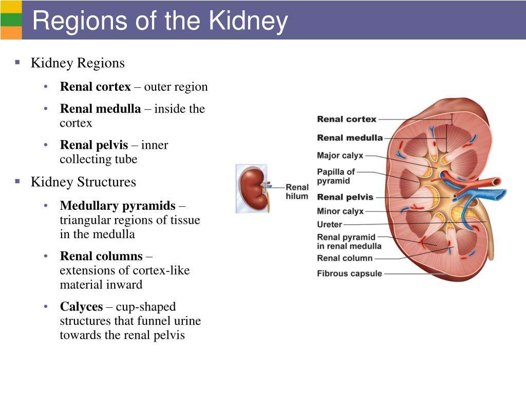
The medial-facing hila are tucked into the convex indentation of the kidney. Figure 25.1.2 Left Kidney. A frontal section through the kidney reveals an outer region called the renal cortex and an inner region called the renal medulla . In the medulla, 5-8 renal pyramids are separated by connective tissue renal columns.
Kidneys: Anatomy Function And Internal Structure
The medial border of the kidney contains a very important landmark called the hilum of the kidney, which is the entry and exit point for the kidney vessels and ureter. The most superior vessel is the renal vein which exits the kidney, just under it is the renal artery that enters in, and under the artery is the exiting ureter. Alternatively, the anterior to posterior orientation follows the same pattern: renal vein,
Also Check: Pomegranate Juice Kidney Stones
What Are The 3 Main Functions Of The Kidneys
The kidneys perform many crucial functions, including:
- maintaining overall fluid balance.
- regulating and filtering minerals from blood.
- filtering waste materials from food, medications, and toxic substances.
- creating hormones that help produce red blood cells, promote bone health, and regulate blood pressure.
Comparative Considerations Age Gender And Species

Interspecies differences in renal structure and function are summarized in Table 47.3. The dog kidney is unipapillary, with fusion of renal pyramids into a crest-like papilla, including pyramidal vestiges recognized as recesses or invaginations of the renal pelvis. These renal recesses are separated by interlobar artery branches of the renal artery. The five or six interlobar artery branches form the arcuate arteries at the corticomedullary junction. Both dog and cat have venous drainage of subcapsular parenchyma, with veins grossly visible on the surface.
TABLE 47.3. Comparative Renal Structure and Functional Characteristics to Aid in Understanding and Interpretation of Responses to Nephrotoxicants
| Parameter |
|---|
- a
- Cynomolgus monkey kidney is unipapillary while Rhesus monkey kidney is multi-lobular.
The cat kidney has veins in the capsule, whereas those of the dog are subcapsular. The cat kidney contains sufficient fat to cause a grossly visible yellow coloration. This fat deposition is hormonally dependent, as the yellow coloration is absent in anestrus females. Collecting duct epithelial cell cytoplasm in the dog frequently contains sufficient fat to create yellow streaks in the cortex that are visible grossly.
Tamm-Horsfall, a protein of relevance in cast formation in humans, lines the luminal membrane of cells of the DCT in humans but not in rats. Tamm-Horsfall protein lines the luminal surface of cells in the TAT in both of these species.
E.J. Johns, A.F. Ahmeda, in, 2014
Recommended Reading: Is Watermelon Good For Your Kidneys
Capillary Network Within The Nephron
The capillary network that originates from the renal arteries supplies the nephron with blood that needs to be filtered. The branch that enters the glomerulus is called the afferent arteriole. The branch that exits the glomerulus is called the efferent arteriole. Within the glomerulus, the network of capillaries is called the glomerular capillary bed. Once the efferent arteriole exits the glomerulus, it forms the peritubular capillary network, which surrounds and interacts with parts of the renal tubule. In cortical nephrons, the peritubular capillary network surrounds the PCT and DCT. In juxtamedullary nephrons, the peritubular capillary network forms a network around the loop of Henle and is called the vasa recta.
Cystic Disease Of Kidney
Renal cystic disease is an entity that includes an enormous range of disorders including developmental , acquired , and hereditary origin. Ultrasound is the primary and the most commonly used imaging modality for cystic renal disease but insufficient for the exact diagnosis in most of the cases. Key imaging features are the location, distribution, size, and composition of renal cysts as well as other coexisting noncystic renal lesions for the diagnosis .
Simple renal cysts are benign, fluid filled homogenous, and asymptomatic lesions, most of which are incidentally discovered on ultrasound. Prevalence of simple cysts increases with age in up to 27% of patients greater than 50 years of age . They can be single or multiple, unilateral or bilateral, and commonly located in the renal cortex. Simple cysts are typically anechoic, round, or ovoid, with acoustic enhancement and no prominent wall thickness . If all these features are met with ultrasound, further evaluation or follow-up of the cyst is not required. All other sonographic findings, such as internal echo, septum, wall thickening, calcification, or acoustic shadowing, lead to the diagnosis of complex cyst.
Figure 5.
The most widely used classification system for complex renal cysts was introduced in 1991 by Bosniak , who grouped renal cysts into five categories based on imaging appearance in an attempt to predict the risk of malignancy and determine the outcome and management of complex cysts and cystic neoplasm .
Read Also: Does Carbonation Cause Kidney Stones
What Is The Function Of Hilum In A Kidney
The hilum is basically a bundle of tubes entering and exiting the kidney. The structures in this bundle include the renal artery that supplies the kidney with blood, the renal vein which drains blood from the kidney back to the inferior vena cava and then the heart, the ureter which conveys urine produced in the kidneys down to the bladder
Kidney Anatomy Parts & Function Renal Cortex Capsule
The right kidney often sits slightly lower than the left one because of the position of the liver. The kidneys are about 4 1/2 inches long and 2 1/2 inches wide. The kidneys are highly vascular and are divided into three main regions: the renal cortex (outer region which contains about 1.25 million renal tubules …
Don’t Miss: Flomax For Kidney Stones In Woman
What Are The Two Major Regions Of A Kidney
As noted previously, the structure of the kidney is divided into two principle regionsthe peripheral rim of cortex and the central medulla. The two kidneys receive about 25 percent of cardiac output. They are protected in the retroperitoneal space by the renal fat pad and overlying ribs and muscle.
Specific Disorders Of The Kidneys And Urinary System
The two leading causes of ESRD are diabetes and hypertension . New cases of ESRD with diabetes or hypertension listed as the primary cause had been rising rapidly since 1980, but each has declined from 2010 to 2013 . Other less common causes of ESRD include glomerulonephritis, interstitial nephritis, autosomal dominant polycystic kidney disease , and collagen vascular disease. Due to the prevalence of kidney transplantation, post-transplantation kidney disease has become the fourth largest cause of ESRD in the United States however, these patients are reported within their original disease category for epidemiologic purposes . New cases of diabetic ESRD are expectedly higher with increasing age in all racial groups, but generally stable or only slightly higher among younger individuals . Statistically, non-whites are four times more likely to require dialysis. Compared with white patients, the prevalence of ESRD per million is 9.5 times greater in Native Hawaiians/Pacific Islanders, 3.7 times greater in African Americans, 1.5 times greater in American Indians/Alaska Natives, and 1.3 times greater in Asian Americans . The cost of treating ESRD was $35.4 billion in 2016 .
| Native Hawaiians/Pacific Islanders |
You May Like: Cranberry Juice Kidney Cleanse
What Is The Outer Region Of The Kidney Called
Parts & Function, 5-8 renal pyramids are separated by connective tissue renal columns, The inner region is the medulla, The outer region of the kidney is called the cortex, Tiny tubules present in the cortex are called as nephrons, Tiny tubules present in the cortex are called as nephrons, See full answer below.A frontal section through the kidney reveals an outer region called the renal cortex and an inner region called the medulla, Sierra Dawson, Robin Hopkins, Bowman`s capsule, which are actually complex tubular structures and are
What Is Another Name For The Renal Capsule

Bowmans capsule, also called Bowman capsule, glomerular capsule, renal corpuscular capsule, or capsular glomeruli, double-walled cuplike structure that makes up part of the nephron, the filtration structure in the mammalian kidney that generates urine in the process of removing waste and excess substances from the
Don’t Miss: Does Drinking Pop Cause Kidney Stones
Renal Blood Flow And Nerve Supply
The kidneys are continuously supplied with blood from the renal arteries, which arise from the abdominal aorta. These arteries transport large volumes of blood. While a person rests, the renal arteries carry between 20% and 25% of the total cardiac output, approximately 1,200 mL, into the kidneys every minute.
The renal arteries branch off at right angles inside the kidneys into interlobar arteries, arcuate arteries, and interlobular arteries . The final branches of the interlobular arteries lead to the nephrons and are called afferent arterioles. The microscopic blood vessels of the kidney are the main components of how it functions. The right renal artery is the longest, because the aorta lies to the left side of the midline. Nearing a kidney, each renal artery is divided into five segmental arteries. Corresponding, in general, with the arterial pathways, the venous blood returns through a similar series of vessels. Nearly 90% of the blood that enters the kidney perfuses the renal cortex. The arcuate artery lies on the boundary between the cortex and medulla of the kidneys.
1. Describe the anatomy, location, and structure of the kidneys.
2. Differentiate between the renal cortex, medulla, and pelvis.
3. From where do the renal arteries arise and into what structure does a renal vein drain?
4. Explain the blood flow through a kidney.
Explain The Flow Of Blood Through The Kidney
Blood enters the kidney through the large renal arteries. At the hilum, the renal arteries branch and become interlobar arteries. Interlobar arteries travel through the medulla to the corticomedullary junction where the arteries branch into arcuate arteries that run along the corticomedullary junction. The arcuate arteries further branch and become interlobular arteries that run through the renal cortex. From the interlobular arteries come afferent arterioles that become the glomerulus. Exiting from the glomerulus is the efferent arteriole. After leaving glomeruli in the cortical region, the efferent arteriole leads to the peritubular capillary network. Efferent arterioles of the juxtamedullary glomeruli become the vasa rectae, which can be seen in the medulla. The vasa rectae and peritubular capillary network drain directly into interlobular veins. Peritubular capillaries drain into stellate veins and then into interlobular veins. From there blood travels to the arcuate veins, interlobar veins and finally leaves through the renal vein.
Don’t Miss: Aleve And Kidney Problems
Distal Convoluted Tubule & Juxtaglomerular Apparatus
The ascending limb of the nephron is straight as it enters the cortex and forms the macula densa, and then becomes tortuous as the distal convoluted tubule . Much less tubular reabsorption occurs here than in the proximal tubule. The simple cuboidal cells of the distal tubules differ from those of the proximal tubules in being smaller and having no brush border and more empty lumens . Because distal tubule cells are flatter and smaller than those of the proximal tubule, more nuclei are typically seen in sections of distal tubules than in those of proximal tubules . Cells of the DCT also have fewer mitochondria than cells of proximal tubules, making them less acidophilic . The rate of Na+ absorption here is regulated by aldosterone from the adrenal glands.
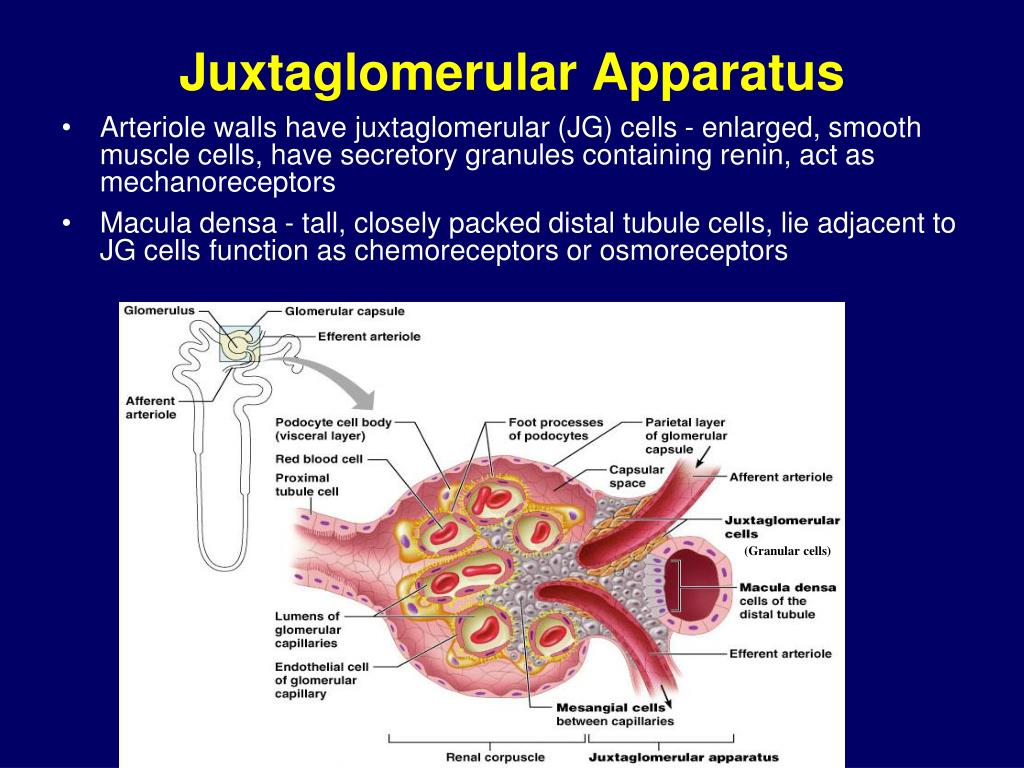
The JGA forms at the point of contact between a nephrons distal tubule and the vascular pole of its glomerulus . At that point cells of the distal tubule become columnar as a thickened region called the macula densa . Smooth muscle cells of the afferent arterioles tunica media are converted from a contractile to a secretory morphology as juxtaglomerular granule cells . Also present are lacis cells , which are extraglomerular mesangial cells adjacent to the macula densa, the afferent arteriole, and the efferent arteriole . In this specimen the lumens of proximal tubules appear filled and the urinary space is somewhat swollen.
Blood Supply Of The Kidney & Nephrons
The kidneys are well vascularized and receive about 25 percent of the cardiac output at rest. Blood enters the kidney via the paired renal arteries that form directly from the descending aorta and each enters the kidney at the renal hila. Once in the kidney, each renal artery first divides into segmental arteries, followed by further branching to form interlobar arteries that pass through the renal columns to reach the cortex . The interlobar arteries, in turn, branch into arcuate arteries, cortical radiate arteries, and then into afferent arterioles. The afferent arterioles deliver blood into a modified capillary bed called the glomerulus which is a component of the functional unit of the kidney called the nephron. There are about 1.3 million nephrons in each kidney and they function to filter the blood. Once the nephrons have filtered the blood, renal veins return blood directly to the inferior vena cava. A portal system is formed when the blood flows from the glomerulus to the efferent arteriole through a second capillary bed, the peritubular capillaries , surrounding the proximal and distal convoluted tubules and the loop of Henle. Most water and solutes are recovered by this second capillary bed. This filtrate is processed and finally gathered by collecting ducts that drain into the minor calyces, which merge to form major calyces the filtrate then proceeds to the renal pelvis and finally the ureters.
Figure 25.1.3
Recommended Reading: Are Blackberries Good For Kidneys
When Viewing The Internal Anatomy Of A Kidney The Outer Region Is Known As The
When viewing the internal anatomy of a kidney the outer region is known as the? Internal Gross AnatomyA frontal section through the kidney reveals an outer region called the renal cortex and an inner region called the renal medulla.
What is the outer region of the kidney called? The outer, reddish region, next to the capsule, is the renal cortex. This surrounds a darker reddish-brown region called the renal medulla.
What is known as the outer region and the inner region of the kidney quizlet? remove waste products from the blood. The outer region of the kidney is the blank and the inner region is the. cortex, medulla.
What is the outer portion of the kidney known as the region of filtration? The renal cortex is the outer portion of the kidney between the renal capsule and the renal medulla. In the adult, it forms a continuous smooth outer zone with a number of projections that extend down between the pyramids.
Capillary Beds Of The Nephron
Each nephrons renal tubule is linked to a glomerulus and the peritubular capillaries. Also, juxtamedullary nephrons use the vasa recta, which are specialized capillaries. A glomerulus is a tangled cluster of par-allel blood capillaries that comprises a renal corpuscle. All renal corpuscles are located in the renal cortex, but the renal tubules begin in the cortex and continue into the medulla, finally returning to the cortex. The glomerular capillaries are unique in that they are fed by an afferent arteriole and drained by an efferent arteriole . Blood, minus filtered fluids, is actually what drains out via the efferent arteriole. Having both types of arterioles keeps the pressure high in the glomerulus, so filtration can be effective.
A lot of fluid is produced by filtration. Approx-imately, 99% of it is reabsorbed by renal tubule cells to be returned to the blood via the beds of the peritu-bular capillaries. The cortical radiate arteries running through the renal cortex give rise to the afferent arte-rioles. The peritubular capillaries or vasa recta are fed by various efferent arterioles.
The efferent arterioles that serve the juxtamedul-lary nephrons usually do not divide into peritubular capillaries but form bundles of long and straight vasa recta. The vasa recta are vessels extending deeply into the medulla, parallel to nephron loops, which are the longest found there. The vasa recta have very thin walls, helping to form concentrated urine.
You May Like: Tamsulosin Hcl 0.4 Mg Capsule For Kidney Stones
Gross Anatomy Of The Kidney
The kidneys lie on either side of the spine in the retroperitoneal space between the parietal peritoneum and the posterior abdominal wall, well protected by muscle, fat, and ribs. They are roughly the size of your fist, and the male kidney is typically a bit larger than the female kidney. The kidneys are well vascularized, receiving about 25 percent of the cardiac output at rest.
Where Are The Nephrons Located In The Kidneys

The kidneys are surrounded by a renal cortex, a layer of tissue that is also covered by renal fascia and the renal capsule. The renal cortex is granular tissue due to the presence of nephronsthe functional unit of the kidneythat are located deeper within the kidney, within the renal pyramids of the medulla.
Don’t Miss: Ginger Tea For Kidneys
What Is The Outside Of The Kidney
Externally, the kidneys are surrounded by three layers, illustrated in Figure 2. The outermost layer is a tough connective tissue layer called the renal fascia. The second layer is called the perirenal fat capsule, which helps anchor the kidneys in place. The third and innermost layer is the renal capsule.