Renal Pelvis And Ureter
Numerous collecting ducts merge into the renal pelvis, which then becomes the ureter. The ureter is a muscular tube, composed of an inner longitudinal layer and an outer circular layer. The lumen of the ureter is covered by transitional epithelium . Recall from the Laboratory on Epithelia that the transitional epithelium is unique to the conducting passages of the urinary system. Its ability to stretch allows the dilation of the conducting passages when necessary. The ureter connects the kidney and the urinary bladder.
The Pain Isn’t Decreasing After More Than A Week
The spleen sits under your rib cage in the upper left part of your abdomen toward your back. Lower left back pain from a kidney stone may be felt when a stone moves inside the left kidney, or moves through the ureters, thin tubes connecting the kidneys . Understanding the nine regions and four quadrants of your abdomen can help. Like kidney stones, gallstones are hard masses that form in the gallbladder and bile ducts. Organs found in the right lower quadrant include the appendix, the upper . Having discomfort in this region, either as right or left flank pain, . If you’re experiencing pain or discomfort in your upper right back area but can’t figure out what’s causing it, don’t worry we’re here to . Flank pain is pain in one side of the body between the upper belly area and the back. Call your healthcare provider right away if . Healthcare professionals refer to this area as the lower . The pain isn’t decreasing after more than a week. Your gallbladder is a small organ on the right side . Middle back pain is any type of pain or discomfort in the area between your upper and lower back.
If you’re experiencing pain or discomfort in your upper right back area but can’t figure out what’s causing it, don’t worry we’re here to . It is an organ that is part of the lymph system . Pain or numbness in your leg Pain in a new area of your back Having discomfort in this region, either as right or left flank pain, .
Blood Supply Of The Kidney & Nephrons
The kidneys are well vascularized and receive about 25 percent of the cardiac output at rest. Blood enters the kidney via the paired renal arteries that form directly from the descending aorta and each enters the kidney at the renal hila. Once in the kidney, each renal artery first divides into segmental arteries, followed by further branching to form interlobar arteries that pass through the renal columns to reach the cortex . The interlobar arteries, in turn, branch into arcuate arteries, cortical radiate arteries, and then into afferent arterioles. The afferent arterioles deliver blood into a modified capillary bed called the glomerulus which is a component of the functional unit of the kidney called the nephron. There are about 1.3 million nephrons in each kidney and they function to filter the blood. Once the nephrons have filtered the blood, renal veins return blood directly to the inferior vena cava. A portal system is formed when the blood flows from the glomerulus to the efferent arteriole through a second capillary bed, the peritubular capillaries , surrounding the proximal and distal convoluted tubules and the loop of Henle. Most water and solutes are recovered by this second capillary bed. This filtrate is processed and finally gathered by collecting ducts that drain into the minor calyces, which merge to form major calyces the filtrate then proceeds to the renal pelvis and finally the ureters.
Figure 25.1.3
Read Also: Coke And Asparagus Recipe For Kidney Stones
What Is Urine Made Of
Urine is made of water, urea, electrolytes, and other waste products. The exact contents of urine vary depending on how much fluid and salt you take in, your environment, and your health. Some medicines and drugs are also excreted in urine and can be found in the urine.
- 94% water
- .1% uric acid
*Electrolytes
As mentioned prior, urine is formed in the nephrons by a three-step process: glomerular filtration, tubular re-absorption, and tubular secretion. The amount of urine varies based on fluid intake and ones environment.
Crossing Vessel Upj Obstruction Vesicoureteral Reflux

Crossing vessel, ureteropelvic junction obstruction, or vesicoureteral reflux can become pathophysiologic if it causes extrinsic or primary intrinsic obstruction leading to hydronephrosis. This can be seen with aberrant crossing vessels in a single system, which leads to UPJ obstruction. Obstruction can also occur from an ectopic ureter, where it is commonly seen inserting inferomedially in an abnormal location and is often associated with the upper pole moiety of a complete duplicated collecting system.
Similarly, a ureterocele in a single system, or sometimes seen in a complete duplicated system, can cause obstruction. From an intrinsic standpoint, UPJO can also be caused by an adynamic/aperistaltic segment of ureter that is due to abnormal embryologic development. Secondary etiologies of obstruction include stones, infections, iatrogenic ureteral damage causing strictures, and other acquired factors that are not due to anatomic variants.
Vesicoureteral reflux is another variation and is caused by an abnormal insertion of the ureter in the bladder in an abnormal position . This insertion site leads to a shorter intramural tunnel length for the ureter to pass through the bladder wall, which leads to inadequate compression of the ureter during bladder filling and contraction and may allow reflux of urine up the ureter. Vesicoureteral reflux can contribute to pyelonephritis and, in extreme situations, irreversible damage to an affected renal unit.
The Nutcracker Syndrome
Recommended Reading: Are Grapes Good For Kidney Stones
What Is Kidney Cancer
A tumor is a mass of abnormally growing cells. Tumors can be either benign or malignant. Benign tumors have uncontrolled cell growth, but without any invasion into normal tissues and without any ability to spread to distant parts of the body. A tumor is malignant, or cancerous, if tumor cells are able to invade tissues and spread locally, as well as to distant parts of the body. Therefore, kidney cancer occurs when cells in either the cortex of the kidney, or cells in the renal pelvis, grow uncontrollably and form tumors that can invade normal tissues and spread to other parts of the body.
Renal cell carcinoma is the most common type of kidney cancer. It accounts for about 9 out of 10 cases of kidney cancer. In renal cell carcinoma, malignant tumors can be growing in either one or both kidneys and there may be multiple tumors. There are several types of renal cell carcinoma. The type is determined by the appearance of the cancer cell under a microscope. Types of renal cell cancer include:
Transitional cell carcinomas, also known as urothelial carcinomas, account for about 5 to 10 out of every 100 diagnoses of kidney cancer. Transitional cell carcinoma is cancer in the lining of the renal pelvis where urine is stored, before it enters the ureter to then travel to the bladder. This type of kidney cancer looks similar to bladder cancer cells when viewed under a microscope.
Kidney Function And Physiology
Kidneys filter blood in a three-step process. First, the nephrons filter blood that runs through the capillary network in the glomerulus. Almost all solutes, except for proteins, are filtered out into the glomerulus by a process called glomerular filtration. Second, the filtrate is collected in the renal tubules. Most of the solutes get reabsorbed in the PCT by a process called tubular reabsorption. In the loop of Henle, the filtrate continues to exchange solutes and water with the renal medulla and the peritubular capillary network. Water is also reabsorbed during this step. Then, additional solutes and wastes are secreted into the kidney tubules during tubular secretion, which is, in essence, the opposite process to tubular reabsorption. The collecting ducts collect filtrate coming from the nephrons and fuse in the medullary papillae. From here, the papillae deliver the filtrate, now called urine, into the minor calyces that eventually connect to the ureters through the renal pelvis. This entire process is illustrated in Figure 22.7.
You May Like: Do Multivitamins Cause Kidney Stones
How Is Kidney Cancer Staged
With these tests, a stage is determined to help decide the treatment plan. The stage of cancer, or extent of disease, is based on information gathered through the various tests done as the diagnosis and work-up of the cancer is being performed.
Kidney cancer is most commonly staged using the TNM system. The TNM system is used to describe many types of cancers. It has three components:
- T-describing the extent of the “primary” tumor .
- N-describing if there is cancer in the lymph nodes.
- M-describing the spread to other organs .
The staging system is very complex. The entire staging system is outlined at the end of this article. Though complicated, the staging system helps healthcare providers determine the extent of the cancer, and in turn, make treatment decisions for a patient’s cancer.
In addition to the TNM system, kidney cancers are assigned a prognostic risk group . This grouping is based on certain factors that may indicate your ability to tolerate treatments. These include time since diagnosis, performance status, certain lab values and the absence or presence of metastasis. These categories can impact treatment options.
Blood Flows In And Out Of The Kidneys Through Renal Arteries And Veins
Blood enters the kidneys through renal arteries. These arteries branch into tiny capillaries that interact with urinary structures inside the kidneys . Here the blood is filtered. Waste is removed and vital substances are reabsorbed back into the bloodstream. The filtered blood leaves through the renal veins. All the blood in the body moves in and out of the kidneys hundreds of times each daythats about 200 quarts of liquid to be filtered every 24 hours.
You May Like: Is Orange Juice Good For Kidney Disease
Left Hand Side Of Back What Organs Are In The Back Region : Love At Palm
Pain or numbness in your leg Middle back pain is any type of pain or discomfort in the area between your upper and lower back. If you’re experiencing pain or discomfort in your upper right back area but can’t figure out what’s causing it, don’t worry we’re here to . Organs found in the right lower quadrant include the appendix, the upper . Your gallbladder is a small organ on the right side .
What Are The Kidneys
The kidneys are two bean-shaped organs. They are located in the back of the abdomen directly in front of where the lowest ribs can be felt on a person’s back. The kidneys have many important functions essential for life, including filtering the blood, removing waste products from the blood, and ensuring that electrolytes in the blood are correctly balanced. These waste products then become urine. In addition, the kidneys produce two important hormones: Erythropoietin, which is responsible for producing red blood cells that carry oxygen throughout the body, and rennin, which helps control blood pressure.
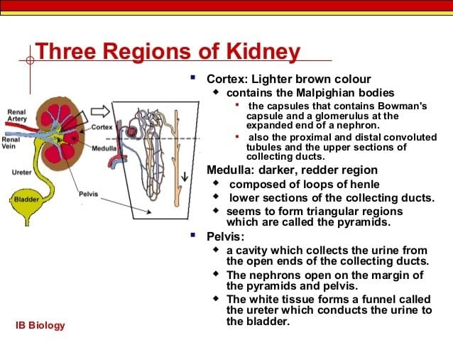
Each of the kidneys can be divided into two main functional parts – the cortex and the renal pelvis. The outer region of the kidney is called the cortex. The cortex consists of a series of tubes and is responsible for filtering blood. The inner region of the kidney is called the renal pelvis. The renal pelvis contains medullary pyramids that collect the filtrate from collecting tubules in the cortex and send it through the ureters to the urinary bladder. Different types of cancers develop in the two different regions of the kidneys.
Recommended Reading: Is Pineapple Good For Kidney Patients
What Are Signs Of Kidney Cancer
The most common sign of kidney cancer is blood in the urine. Blood in the urine may be seen by the naked eye or found only when the urine is analyzed in a laboratory .
Other common symptoms of kidney cancer include:
- Pain in the low back, usually on one side.
- A mass you or your healthcare provider can feel on your side or lower back.
- Feeling tired.
- Fever.
- Anemia .
Symptoms caused by metastatic disease include fever, weight loss, and night sweats . Other symptoms include hypertension, increased calcium in the blood, and liver problems.
The Kidneys Are Retroperitoneal Organs In The Abdomen

The kidneys are located behind the peritoneum, and so are called retroperitoneal organs. They sit in the back of the abdomen between the levels of the T12 and L03 vertebrae. The right kidney is slightly lower than the left kidney to accommodate the liver. Both kidneys are bean-shaped and about the size of an adult fist.
Also Check: Kidney Failure Symptoms In Men
Nephrons: The Basic Functional Units Of Blood Filtration And Urine Production
Each kidney contains over 1 million tiny structures called nephrons. The nephrons are located partly in the cortex and partly inside the renal pyramids, where the nephron tubules make up most of the pyramid mass. Nephrons perform the primary function of the kidneys: regulating the concentration of water and other substances in the body. They filter the blood, reabsorb what the body needs, and excrete the rest as urine.
What Are The Symptoms Of Glomerular Disease
The signs and symptoms of glomerular disease include
- albuminuria: large amounts of protein in the urine
- hematuria: blood in the urine
- reduced glomerular filtration rate: inefficient filtering of wastes from the blood
- hypoproteinemia: low blood protein
- edema: swelling in parts of the body
One or more of these symptoms can be the first sign of kidney disease. But how would you know, for example, whether you have proteinuria? Before seeing a doctor, you may not. But some of these symptoms have signs, or visible manifestations:
- Proteinuria may cause foamy urine.
- Blood may cause the urine to be pink or cola-colored.
- Edema may be obvious in hands and ankles, especially at the end of the day, or around the eyes when awakening in the morning, for example.
Also Check: Is Wine Bad For Kidney Stones
Kidney Disease And Disorders
Kidney diseases and kidney problems are usually treated by a nephrologist. Kidney stones are sometimes treated by a urologist. Here is a list of some of the more common kidney problems:
- Glomerulonephritis inflammation of the glomeruli
- Hydronephrosis excessive fluid within the kidney caused by blocked urine flow
- Pyelonephritis infection of the kidney
- Kidney Stones usually form in the kidneys, but can form anywhere in the urinary tract
- Kidney Cancer
- Nephrosis a process that can lead to kidney failure
- Polycystic Kidney Disease a disorder of the kidneys that result in multiple fluid filled cysts within the kidneys tissues
- Renal Hypertension if the kidneys for some reason do not get enough blood, they set off a series of events leading to high blood pressure
- Renal Infarction similar to a heart attack, but in the kidney, caused by blockage of kidney vessels
- Renal Vein clot clot in the vein that carries blood from the kidney, can be fatal
Capillary Network Within The Nephron
The capillary network that originates from the renal arteries supplies the nephron with blood that needs to be filtered. The branch that enters the glomerulus is called the afferent arteriole. The branch that exits the glomerulus is called the efferent arteriole. Within the glomerulus, the network of capillaries is called the glomerular capillary bed. Once the efferent arteriole exits the glomerulus, it forms the peritubular capillary network, which surrounds and interacts with parts of the renal tubule. In cortical nephrons, the peritubular capillary network surrounds the PCT and DCT. In juxtamedullary nephrons, the peritubular capillary network forms a network around the loop of Henle and is called the vasa recta.
Recommended Reading: Can Kidney Stones Cause Constipation Or Diarrhea
Tubular Reabsorption And Secretion
Tubular reabsorption occurs in the PCT part of the renal tubule. Almost all nutrients are reabsorbed, and this occurs either by passive or active transport. Reabsorption of water and some key electrolytes are regulated and can be influenced by hormones. Sodium is the most abundant ion and most of it is reabsorbed by active transport and then transported to the peritubular capillaries. Because Na+ is actively transported out of the tubule, water follows it to even out the osmotic pressure. Water is also independently reabsorbed into the peritubular capillaries due to the presence of aquaporins, or water channels, in the PCT. This occurs due to the low blood pressure and high osmotic pressure in the peritubular capillaries. However, every solute has a transport maximum and the excess is not reabsorbed.
In the loop of Henle, the permeability of the membrane changes. The descending limb is permeable to water, not solutes the opposite is true for the ascending limb. Additionally, the loop of Henle invades the renal medulla, which is naturally high in salt concentration and tends to absorb water from the renal tubule and concentrate the filtrate. The osmotic gradient increases as it moves deeper into the medulla. Because two sides of the loop of Henle perform opposing functions, as illustrated in Figure 22.8, it acts as a countercurrent multiplier. The vasa recta around it acts as the countercurrent exchanger.
Kidneys: The Main Osmoregulatory Organ
The kidneys, illustrated in Figure 22.4, are a pair of bean-shaped structures that are located just below and posterior to the liver in the peritoneal cavity. The adrenal glands sit on top of each kidney and are also called the suprarenal glands. Kidneys filter blood and purify it. All the blood in the human body is filtered many times a day by the kidneys these organs use up almost 25 percent of the oxygen absorbed through the lungs to perform this function. Oxygen allows the kidney cells to efficiently manufacture chemical energy in the form of ATP through aerobic respiration. The filtrate coming out of the kidneys is called urine.
You May Like: Can Kidney Stones Make You Constipated
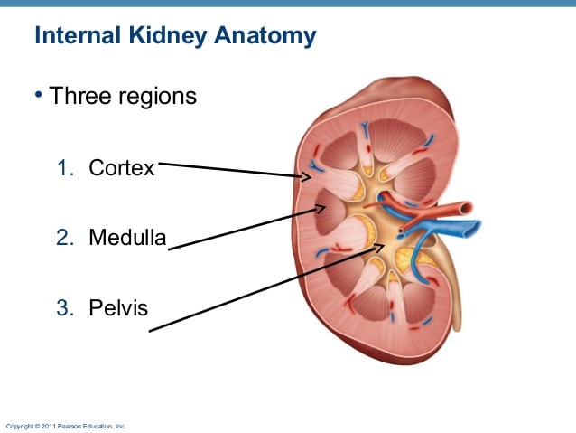
What Are The Regions Of The Kidney
cortex, medulla, and pelvis.The substance, or parenchyma, of the kidney is divided into two major structures: superficial is the renal cortex and deep is the renal medulla.cortex and medulla
Registered users can ask questions, leave comments, and earn points for submitting new answers.
Already have an account? Log in
Ask questions, submit answers, leave comments
Earn points for using the site
Already have an account? Log in