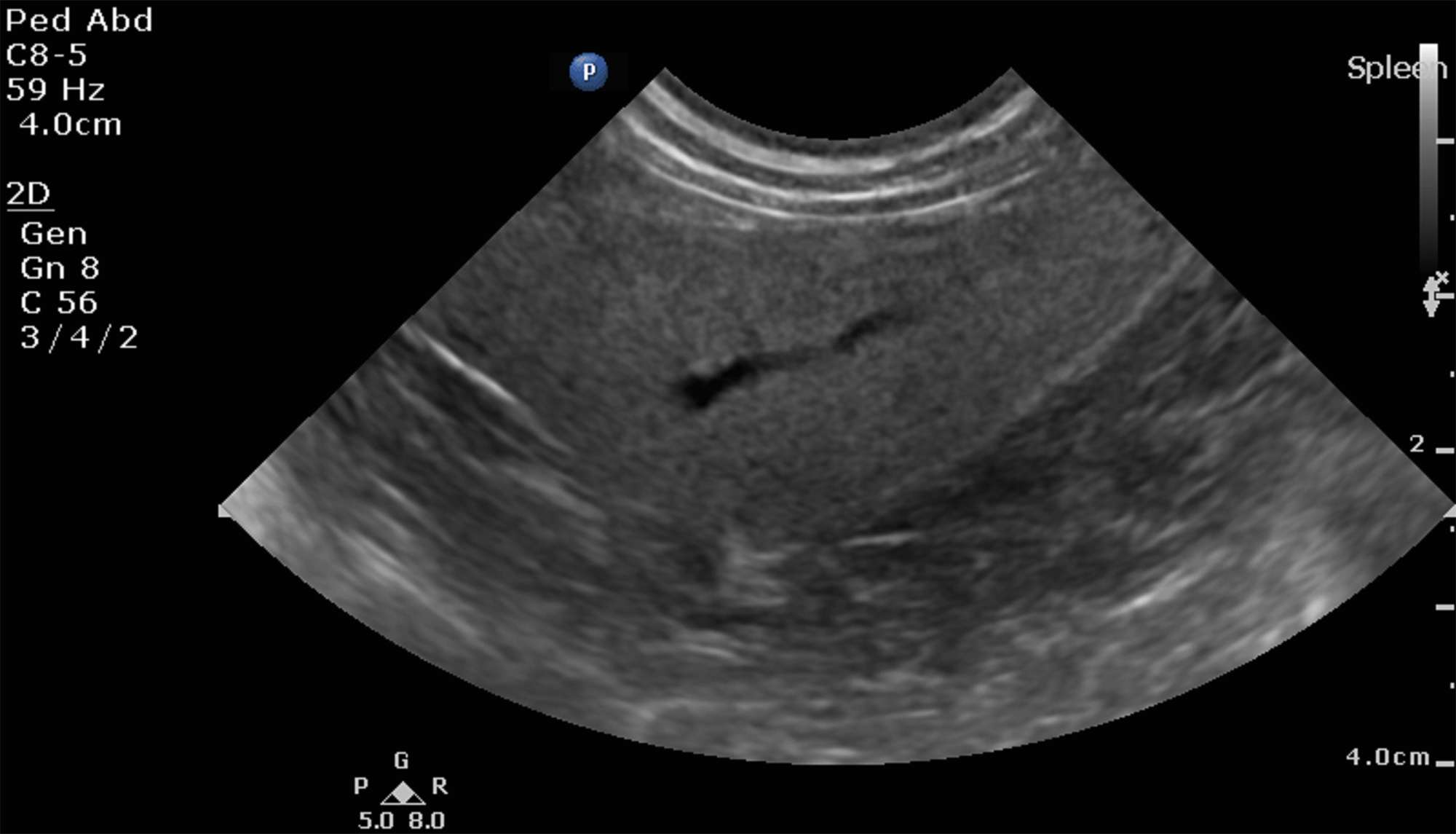
Ultrasound Is Effective In Diagnosing Patients With Kidney Stones
For patients with large kidney stones renal ultrasound is essentially equivalent to CT scan in diagnosis of kidney stones. CT scan is certainly useful for surgical planning in patients with large stones to define anatomy and decide which approach would be ideal especially when the question is whether the patient needs PCNL.
Dont Miss: Can Mio Cause Kidney Stones
Why The Test Is Performed
A pelvic ultrasound is used during pregnancy to check the baby.
A pelvic ultrasound also may be done for the following:
- Cysts, , or other growths or masses in the pelvis found when your doctor examines you
- Bladder growths or other problems
- Kidney stones
- , an infection of a womanâs uterus, ovaries, or tubes
- Abnormal vaginal bleeding
- , a pregnancy that occurs outside the uterus
- Pelvic pain
Why Is A Bladder Ultrasound Done
About a quarter of all people in the United States experience some level of incontinence, or the inability to hold urine in the bladder until you purposely release it.
There are many causes of incontinence, and it can be difficult for a doctor to pinpoint a reason for the problem just by asking you questions or examining the outside of your body.
The following symptoms may lead a doctor to order a bladder ultrasound:
- difficulty urinating
Read Also: Why Is My Right Kidney Sore
What Is Examined During A Renal/pelvic Ultrasound
During a renal/pelvic ultrasound, the kidneys are examined to determine their size, shape and exact position. The bladder may be evaluated to help determine the cause of unexplained blood in the urine or difficulty in urinating, or to look for bladder stones.
Before the renal or pelvic ultrasound
You do not have to fast for this test, but your bladder must be full. See instructions below.
- Finish drinking one quart of fluids 1 hour before your scheduled test. Once you start drinking, do not empty your bladder until the exam is completed.
- Failure to follow the above preparation will result in delays or possible cancellation of your examination.
On the day of the test
Please do not bring valuables such as jewelry and credit cards.
- It is very important to arrive for the test with a full bladder. This allows the technologists and radiologist to view the bladder while it is full and after it has been emptied.
- Your ultrasound test is performed by registered, specially trained technologists, and interpreted by a board-certified radiologist.
- You may be asked to change into a hospital gown.
During the test
- You will lie on a padded examining table.
- A warm, water-soluble gel is applied to the skin over the area to be examined. The gel does not harm your skin or stain your clothes.
- A probe is gently applied against the skin. You may be asked to hold your breath briefly several times.
The ultrasound takes about 40 minutes to complete.
After the test
Next Steps If A Kidney Stone Is Found

If a kidney stone is small enough, it can move or “pass” through your urinary tract and out of your body on its own. If the stone cannot pass on its own, you may need treatment.
Large stones can get stuck in either a kidney or a ureter. A stone that becomes stuck may cause pain that does not go away and may damage the kidney if it is not treated.
If the emergency doctor thinks the kidney stone will pass on its own without any problems:
- You will probably be able to go home.
- You may be given medicines for pain and nausea to take home.
- You may be asked to drink water to help the kidney stone pass.
- You will be asked to watch for the kidney stone when you urinate. You may be told how to strain your urine to catch a stone that passes. If the stone does not pass, call your health care professional.
If the emergency doctor thinks the kidney stone will not pass on its own or may cause problems:
- You may need to stay in the hospital for treatment.
- You may need to see a specialist and may need surgery to remove the stone.
If your nausea and vomiting do not stop:
You may need to stay in the hospital.
You May Like: Can You Improve Kidney Function
How Do I Get Ready For A Kidney Ultrasound
- Your healthcare provider will explain the procedure to you. Ask any questions you have about the procedure.
- You may be asked to sign a consent form that gives permission to do the test. Read the form carefully and ask questions if anything is not clear.
- You usually dont need to stop eating or drinking before the test. You also usually will not need medicine to help you relax .
- The gel put on your skin during the test does not stain clothing, but you may want to wear older clothing. The gel may not be completely removed from your skin afterward.
- You should not empty your bladder before the test if the healthcare provider is going to look at the bladder.
- Follow any other instructions your healthcare provider gives you to get ready.
Detecting Kidney And Urinary Tract Abnormalities Before Birth
Ultrasound examinations are often done as part of prenatal care. This test allows the doctor to examine babies before they are born. With ultrasound, the doctor can see the baby’s internal organs, including the kidneys and urinary bladder. Occasionally, an abnormality is detected in the developing urinary tract. A doctor can then determine whether treatment is necessary. Parents should know that, in many cases, these abnormalities do not have a major impact on the child’s overall health.
You May Like: Is Iga Nephropathy A Chronic Kidney Disease
Preparing For Your Scan
Check your appointment letter for any instructions about how to prepare for your scan.
You usually need to drink about 1 litre of fluid an hour before the test, so that your bladder is comfortably full. Do not empty your bladder before the test. This is so your bladder can be seen clearly in the scan.
Take your medicines as normal unless your doctor tells you otherwise.
How Do I Prepare For My Exam
- Take all prescribed medications as directed.
- Please empty your bladder 90 minutes prior to your appointment, and then drink one litre of water within the next 30 minutes. Finish drinking water one hour before your exam and do not empty your bladder.
- Please note: Drink water slowly to prevent abdominal discomfort.
- If you are too uncomfortable, please relieve your bladder of a small amount of urine.
- Once the test has begun, your sonographer will advise you if you can empty your bladder further or totally. We understand that a full bladder can be uncomfortable.
- Please arrive 15 minutes before your appointment to allow enough time to check in with reception.
- Bring photo identification and your provincial health card.
- Wear comfortable clothes.
- Please dont bring children that require supervision.
Recommended Reading: How Do You Donate A Kidney
What Is A Bladder Ultrasound
A bladder ultrasound is done when a doctor needs to closely examine the structure or function of your bladder.
The bladder is a muscular sac that receives urine from your kidneys, stretching to hold the fluid until you release it during urination. Bladder control, or your ability to control these muscles, makes urination a planned and purposeful task.
However, there are many issues that can complicate the process of urination.
Removing A Stone With Sound Waves
|
Kidney stones can sometimes be broken up by sound waves produced by a lithotriptor in a procedure called extracorporeal shock wave lithotripsy . After an ultrasound device or fluoroscope is used to locate the stone, the lithotriptor is placed against the back, and the sound waves are focused on the stone, shattering it. Then the person drinks fluids to flush the stone fragments out of the kidney, to be eliminated in the urine. Sometimes blood appears in the urine or the abdomen is bruised after the procedure, but serious problems are rare. |
A ureteroscope can be inserted into the urethra, through the bladder and up the ureter to remove small stones in the lower part of the ureter that require removal. In some instances, the ureteroscope can also be used with a device to break up stones into smaller pieces that can be removed with the ureteroscope or passed in the urine . Most commonly, holmium laser lithotripsy is used. In this procedure, a laser is used to break up the stone.
Percutaneous nephrolithotomy may be used to remove some larger kidney stones. In percutaneous nephrolithotomy, doctors make a small incision in the persons back and then insert a telescopic viewing tube into the kidney. Doctors insert a probe through the nephroscope to break the stone into smaller pieces and then remove the pieces .
Making the urine more alkaline may sometimes gradually dissolve uric acid stones. Other types of stones cannot be dissolved this way.
You May Like: What Is Better For The Kidneys Tylenol Or Ibuprofen
What To Expect At A Kidney Ultrasound
A kidney ultrasound is a short, noninvasive procedure. It typically takes about 20 to 30 minutes to complete and involves the following steps:
What Are Kidney And Bladder Stones

Kidney or bladder stones are solid build-ups of crystals made from minerals and proteins found in urine. Bladder diverticulum, enlarged prostate, neurogenic bladder and urinary tract infection can cause an individual to have a greater chance of developing bladder stones.
If a kidney stone becomes lodged in the ureter or urethra, it can cause constant severe pain in the back or side, vomiting, hematuria , fever, or chills.
If bladder stones are small enough, they can pass on their own with no noticeable symptoms. However, once they become larger, bladder stones can cause frequent urges to urinate, painful or difficult urination and hematuria.
Donât Miss: What Organ System Does The Kidney Belong To
Don’t Miss: What Is The Treatment For Kidney Stones
What Happens After Your Imaging Test
After most imaging tests, you can go home and resume normal activity. Some tests that involve catheters may cause minor discomfort. Tests that include medication, dyes, or sedatives occasionally trigger allergic reactions.
Tests that may cause discomfort include
- Tests involving a catheter in the urethra. You might feel some mild discomfort from an irritated urethra for a few hours after the procedure.
- Transrectal ultrasound. You might feel some discomfort from an irritated rectum.
If you have a catheterization, your health care professional may prescribe an antibiotic for 1 or 2 days to prevent an infection. If you have any signs of infection, including pain, chills, or fever, call your health care professional immediately.
Tests that may cause an allergic reaction include
- Tests involving contrast medium. If you have a rare sign of reaction, such as hives, itching, nausea, vomiting, headache, or dizziness, call your health care professional immediately.
- Tests involving sedatives. If you have a rare sign of reaction, such as changes in breathing and heart rate, call your health care professional immediately.
How Should I Prepare For The Scan
You should arrive for your scan with a filled bladder. We recommend that you drink 2 glasses of water a half hour before your scan time. Remember to bring your referral form with you. We will scan you across your tummy to start with and then we will send you to the toilet to empty your bladder before re-scanning you again.
Also Check: Can You See Kidney Stones With Ultrasound
What Are The Risks Of A Kidney Ultrasound
There is no radiation used and generally no discomfort from the applicationof the ultrasound transducer to the skin.
There may be risks depending upon your specific medical condition. Be sureto discuss any concerns with your physician prior to the procedure.
Certain factors or conditions may interfere with the results of the test.These include, but are not limited to, the following:
-
Severe obesity
What Does An Ultrasound Of The Kidneys Show With Certain Specific Medical Issues
Your doctor will perform this procedure if he or she suspects that the organ is not functioning properly in any way. The ultrasound thats performed will need to use high-quality equipment in order to ensure an accurate result. Here are a few examples of signs and symptoms that will lead to your doctor performing one:
- Lack of sufficient urine output
- Unusual pain in the lower back
- Nausea, vomiting, and dizziness
You May Like: How To Reduce Inflammation In Kidneys
Checking For Kidney Stones In The Emergency Department
First, the emergency doctor will give you medicine to help stop your pain. The medicine may be given by mouth. Or, it may be given through an intravenous needle placed in a vein in your arm. You may also be given medicine to help stop your nausea and vomiting. If you are dehydrated from vomiting, you may be given liquids through an IV tube.
Next, the emergency doctor will talk with you about your symptoms and medical history. If the emergency doctor thinks you might have a kidney stone, several tests may be done.
These may include:
- Urine Tests: To check for blood or mineral crystals in your urine or for signs of infection.
- Blood Tests: To check the health of your kidneys and for signs of a kidney or blood infection.
- Imaging Tests: To check for kidney stones in your urinary tract . Imaging tests may include a CT scan or an ultrasound.
How Should I Prepare
Your doctor will give you instructions before the exam. You may have to drink 24 ounces of water before the exam to get better images of your bladder. They may also tell you not to eat or drink hours before the exam. Your doctor may ask you not to urinate until the exam is complete.
Wear comfortable, loose-fitting clothing. You may need to remove all clothing and jewelry in the area under examination. You may need to change into a gown for the procedure.
Ultrasound exams are sensitive to motion, and an active or crying child can prolong the exam. To ensure a smooth experience, it often helps to explain the procedure to the child before the exam. Bring books, small toys, music, or games to help distract the child and make the time pass quickly. The exam room may have a television.
Don’t Miss: Can Metformin Affect Kidney Function
What To Expect After A Kidney Ultrasound
Youll be able to eat and drink as usual after your ultrasound. Additionally, you can return to your daily activities after you leave the facility.
After the ultrasound, the technician will forward the results to a radiologist. This is a type of doctor that specializes in making sense of medical images, such as those created by ultrasound.
Once the radiologist has reviewed your images, which typically takes only 1 or 2 days, theyll send their findings to your doctor. Your doctor will then contact you to go over the results of your ultrasound.
What Happens During A Bladder Ultrasound

In some facilities, you may need to see a special technician for an ultrasound. But some medical offices can do this test in the examination room during a routine appointment.
Whether you have the test done in an examination room or an imaging center, the process will be similar:
Don’t Miss: Is Stevia Bad For Kidneys
What Happens After My Exam
Your images will be reviewed by a specialized radiologist who will compile a report that is sent to your doctor within 24 hours, sooner for urgent requests. Mayfair Diagnostics is owned and operated by over 60 radiologists who are fellowship-trained in many keys areas, such as neuroradiology, body, cardiac, and musculoskeletal imaging, etc. This allows for an expert review of your imaging by the applicably trained radiologist.
Your images will be uploaded to a provincial picture archiving and communication system this technology provides electronic storage and convenient access to your medical images from multiple sources, such as your doctor, specialists, hospitals, and walk-in clinics.
Your doctor will review your images and the report from the radiologist and discuss next steps with you, such as a treatment plan or the need for further diagnostic imaging or lab tests to ensure an accurate diagnosis.
Mayfair Diagnostics has 12 locations across Calgary which provide ultrasound services, as well as one in Cochrane and one in Regina. For more information about our clinic locations and services, please visit our clinic location pages, or you can drop by the nearest clinic.
REFERENCES