What Happens On The Day
When you arrive for your scan you will be asked to fill out a form which helps us understand your concerns.
The sonographer will then take you through to the scanning room.
You will be asked to lie on the table and expose your tummy. A towel will be tucked into your pants to limit spread of the gel onto your clothes.
Clear gel is applied to your tummy and the sonographer moves the probe over your tummy recording images. Measurements will be taken of your bladder whilst it is full and your kidneys.
You will then be sent to the toilet to empty your bladder.
You will be re-scanned after emptying your bladder to see if your bladder has emptied completely.
The sonographer will leave the room after the scan to review the images and to discuss them with the Radiologist. Sometimes for clarification more images are required.
Where Is A Transvaginal Ultrasound Done
The examination is carried out in a radiology department of a hospital, private radiology practice or at a specialist clinic for obstetric and gynaecological imaging. The examination is carried out in the privacy of an ultrasound room, which might be dimly lit to allow the images on the ultrasound screen to be clearly seen. To maintain your privacy, the examination door is closed and there is a sign on the outside stating that an ultrasound examination is in progress.
Why Do I Have Pain In My Left Ovary
According to VeryWellhealth.com, ovary pain, which is often felt in the lower abdomen, pelvis, or lower back, are related to ovulation and menstruation. A GYN problem like endometriosis or pelvic inflammatory disease, or even a medical condition affecting your digestive or urinary system can be to blame.
You May Like: Can Chocolate Cause Kidney Stones
Calyceal Diverticulum Or Calyceal Cyst
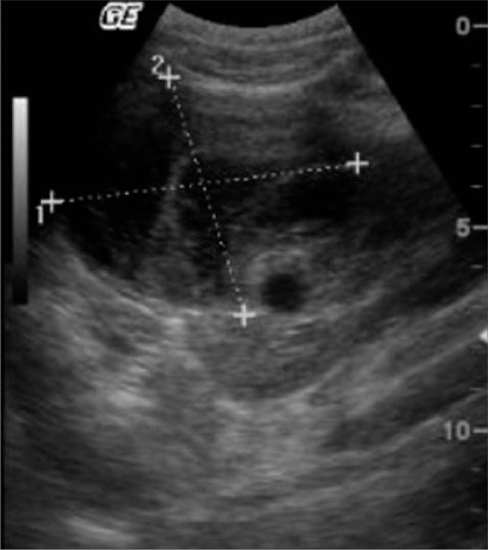
The above B-mode ultrasound image and Color Doppler image show a cystic lesion involving the collecting system of the right kidney. This suggests a calycealdiverticulum /calyceal cyst of the right kidney. The walls of the cystic lesion are thin but irregular and possibly communicate with the renal pelvis. Again, in this case too, the cyst appears todisplace blood vessels which surround the lesion.
When Can I Expect The Results Of My Transvaginal Ultrasound

The time it takes for your doctor to receive a written report will vary. The private radiology practice, clinic or hospital where your procedure is carried out will be able to tell you when your doctor is likely to receive the report.
It is important that you discuss the results with your doctor, either in person or on the telephone, so that they can explain what the results mean for you.
*The author has no conflict of interest with this topic.
Page last modified on 31/8/2017.
Read Also: Is Cranberry Juice Good For Your Liver And Kidneys
Cross Fused Ectopic Kidneys With Duplex Ureters
This male patient shows absence of both right and left kidneys in their normal positions- that is the renal fossae. Both kidneys appear as a single mass in the right iliac fossa- a clear case ofcross fused ectopia of the kidneys. Such a kidney is called a pancake kidney also. These ultrasound images show a typical case of cross fused ectopia of the kidneys with an added anomaly also-there is bilateral duplication of the ureters. The urinary bladder shows 2 ureteric jets- suggesting duplication of the collecting system and ureters, which are seen to open on either side of theurinary bladder.
These ultrasound images of the cross fused renal ectopia with bilateral duplication of the collecting system are courtesy of Dr Ravi Kadasne, MD.
Ultrasound Of The Kidneys
In medicine, KUB refers to a diagnostic medical imaging technique of the abdomen and stands for Kidneys, Ureters, and Bladder, although in fact the Ureters only show if they are abnormally distended. A KUB ultrasound is an examination requested by your doctor to evaluate the urinary tract . In the male patient, the prostate gland is also scanned.
A KUB Ultrasound may be requested:
- To look for changes in the bladder wall
- To look for changes in the kidney size or structure
- To look for stones in the urinary tract
- To evaluate reasons why you have recurrent kidney infection
- To identify the cause of renal or pelvic pain
What happens during my renal or KUB ultrasound?
The urinary bladder can only be properly assessed when full or distended, as bladder volume measurements are taken whilst the bladder is full.
The bladder and both kidneys are also scanned after the bladder has been emptied to evaluate the volume of urine retained
In the male patient the prostatic volume is measured.
During the ultrasound examination we will make a detailed study of the size, shape and condition of the:
- Urinary Bladder, Both full and immediately after emptying
- Both Right and Left Kidneys and their ureters
- The Prostate gland
A written report of the findings will be provided, with images where necessary to demonstrate abnormal findings.
What preparation is required for the a KUB Ultrasound scan?
What will happen during the examination?
Recommended Reading: Renal Diet Orange Juice
What Happens After A Pelvic Ultrasound
There is no special type of care required after a pelvic ultrasound.You may resume your normal diet and activity unless your doctor advisesyou differently.
There are no confirmed adverse biological effects on patients orinstrument operators caused by exposures to ultrasound at the intensitylevels used in a diagnostic ultrasound.
Your doctor may give you additional or alternate instructions after theprocedure, depending on your particular situation.
What Is Ultrasound Imaging
An ultrasound exam is a painless diagnostic technique that makes use of how sound waves travel through the body. When sound waves pass through the body, they bounce off tissues and organs in certain ways. The reflected waves can be used to make images of the organs inside. The sound waves dont hurt the body, and theres no radiation.
Ultrasound imaging may be done in the health providers office, in the hospital, or in an outpatient facility. The reason for the study and details of the case will help decide where the test should be done.
In most cases, very little needs to be done before an ultrasound exam. The patient lies on the exam table. A clear, water-based gel is put on the skin over the part to be checked. This gel helps the sound waves go through the body. A hand-held probe is then moved over that part. For prostate ultrasound exams, a specially designed probe is inserted into the rectum.
There is no risk of radiation. The patient can return to daily tasks right away after the test.
Some exams, such as a bladder scan for residual urine, dont call for the user to have a lot of experience. Other exams, such as ultrasound of the kidneys, testicles or prostate, call for the user to have more experience or skill.
Also Check: Is Pineapple Good For Kidney Stones
What Happens During A Kidney Ultrasound
A kidney ultrasound may be performed on an outpatient basis or as part ofyour stay in a hospital. Although each facility may have differentprotocols in place, generally an ultrasound procedure follows this process:
You will be asked to remove any clothing, jewelry, or other objects that may interfere with the scan.
If asked to remove clothing, you will be given a gown to wear.
You will lie on an examination table on your stomach.
Ultrasound gel is placed on the area of the body that will undergo the ultrasound examination.
Using a transducer, a device that sends out the ultrasound waves, the ultrasound wave will be sent through that patient’s body.
The sound will be reflected off structures inside the body, and the ultrasound machine will analyze the information from the sound waves.
The ultrasound machine will create images of these structures on a monitor. These images will be stored digitally.
If the bladder is examined, you will be asked to empty your bladder after scans of the full bladder have been completed. Additional scans will be made of the empty bladder.
There are no confirmed adverse biological effects on patients or instrumentoperators caused by exposures to ultrasound at the intensity levels used indiagnostic ultrasound.
What Medical Problems Can Be Diagnosed With A Pelvic Ultrasound
The most common reason for a bladder ultrasound is to assess the bladder wall, bladder capacity and post-void residual . Bladder ultrasound can detect bladder stones, bladder tumors and bladder diverticula. It may also detect ureteroceles among other urological problems.
A pelvic ultrasound can help identify bladder tumors, kidney stones, and other disorders of the urinary tract in both men and women.
You May Like: Carbonation And Kidney Stones
Findings In The Normal Kidney
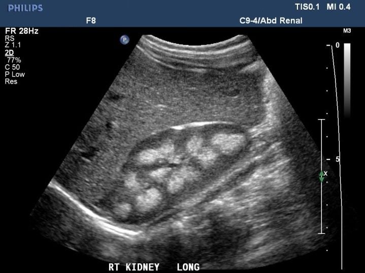
In the longitudinal scan plane, the kidney has the characteristic oval bean-shape. The right kidney is often found more caudally and is slimmer than the left kidney, which may have a so-called dromedary hump due to its proximity to the spleen . The kidney is surrounded by a capsule separating the kidney from the echogenic perirenal fat, which is seen as a thin linear structure .
The kidney is divided into parenchyma and renal sinus. The renal sinus is hyperechoic and is composed of calyces, the renal pelvis, fat and the major intrarenal vessels. In the normal kidney, the urinary collecting system in the renal sinus is not visible, but it creates a heteroechoic appearance with the interposed fat and vessels. The parenchyma is more hypoechoic and homogenous and is divided into the outermost cortex and the innermost and slightly less echogenic medullary pyramids . Between the pyramids are the cortical infoldings, called columns of Bertin . In the pediatric patient, it is easier to differentiate the hypoechoic medullar pyramids from the more echogenic peripheral zone of the cortex in the parenchyma rim, as well as the columns of Bertin .
Normal pediatric kidney. * Column of Bertin ** pyramid *** cortex **** sinus.
Measures of the kidney. L = length. P = parenchymal thickness. C = cortical thickness.
What Is Examined During A Renal/pelvic Ultrasound

During a renal/pelvic ultrasound, the kidneys are examined to determine their size, shape and exact position. The bladder may be evaluated to help determine the cause of unexplained blood in the urine or difficulty in urinating, or to look for bladder stones.
Before the renal or pelvic ultrasound
You do not have to fast for this test, but your bladder must be full. See instructions below.
- Finish drinking one quart of fluids 1 hour before your scheduled test. Once you start drinking, do not empty your bladder until the exam is completed.
- Failure to follow the above preparation will result in delays or possible cancellation of your examination.
On the day of the test
Please do not bring valuables such as jewelry and credit cards.
- It is very important to arrive for the test with a full bladder. This allows the technologists and radiologist to view the bladder while it is full and after it has been emptied.
- Your ultrasound test is performed by registered, specially trained technologists, and interpreted by a board-certified radiologist.
- You may be asked to change into a hospital gown.
During the test
- You will lie on a padded examining table.
- A warm, water-soluble gel is applied to the skin over the area to be examined. The gel does not harm your skin or stain your clothes.
- A probe is gently applied against the skin. You may be asked to hold your breath briefly several times.
The ultrasound takes about 40 minutes to complete.
After the test
Read Also: Is Grape Juice Good For Kidney Stones
How The Test Is Performed
During the procedure, you will lie on your back on the table. Your health care provider will apply a clear gel on your abdomen.
Your provider will place a probe , over the gel, rubbing back and forth across your belly:
- The probe sends out sound waves, which go through the gel and reflect off body structures. A computer receives these waves and uses them to create a picture.
- Your provider can see the picture on a TV monitor.
Depending on the reason for the test, women also may have a transvaginal ultrasound during the same visit.
How Do I Prepare For My Pelvic Ultrasound Appointment
- Before your ultrasound, your doctor will explain how the scan will work. If you have any questions, be sure to ask.
- If you are sensitive or allergic to latex, be sure to tell your doctor before your ultrasound.
- Most women can eat and drink before their scan. You wont need any medicine to help you relax or go to sleep. Your doctor may give you medication if your ultrasound is part of another procedure that uses anesthesia.
- Wear clothing you can get gel on. The gel that technicians put on your skin wont stain your clothes, but some of the gel may stick to your skin after your ultrasound.
- If youre having a transabdominal ultrasound, your provider will ask you to drink several glasses of water one to two hours before your ultrasound. Be sure not to go to the bathroom and urinate until after your ultrasound is over.
- If youre having a transvaginal ultrasound, be sure to urinate right before your ultrasound.
- Follow all other directions from your doctor before the procedure.
Recommended Reading: Can Seltzer Water Cause Kidney Stones
How Do I Prepare For A Kidney Ultrasound
EAT/DRINK: Drink a minimum of 24 ounces of clear fluid at least one hour before yourappointment. Do not empty your bladder prior to the procedure. Generally,no prior preparation, such as fasting or sedation, is required.
Your physician will explain the procedure to you and offer you theopportunity to ask any questions that you might have about the procedure.
You may be asked to sign a consent form that gives your permission to dothe procedure. Read the form carefully and ask questions if something isnot clear.
Based upon your medical condition, your physician may request otherspecific preparation.
When Should I Know The Results Of An Abdominal Ultrasound Test
After your test, a radiologist reviews the ultrasound pictures. This medical expert writes a report of the test findings and sends it to your provider. You should hear about your results from your provider within one week.
Providers sometimes use ultrasound to diagnose potentially life-threatening problems in an emergency. If your provider suspects an urgent concern, you will get results right away.
Don’t Miss: Is Watermelon Good For Your Kidneys
How Do I Prepare For A Pelvic Ultrasound
EAT/DRINK: Drink a minimum of 24 ounces of clear fluid at least one hour beforeyour appointment. Do not empty your bladder until after the exam.
Generally, no fasting or sedation is required for a pelvic ultrasound,unless the ultrasound is part of another procedure that requiresanesthesia.
For a transvaginal ultrasound, you should empty your bladder rightbefore the procedure.
Your doctor will explain the procedure to you and offer you theopportunity to ask any questions that you might have about theprocedure.
Based on your medical condition, your doctor may request other specificpreparation.
What Are Kidney And Bladder Stones
Kidney or bladder stones are solid build-ups of crystals made from minerals and proteins found in urine. Bladder diverticulum, enlarged prostate, neurogenic bladder and urinary tract infection can cause an individual to have a greater chance of developing bladder stones.
If a kidney stone becomes lodged in the ureter or urethra, it can cause constant severe pain in the back or side, vomiting, hematuria , fever, or chills.
If bladder stones are small enough, they can pass on their own with no noticeable symptoms. However, once they become larger, bladder stones can cause frequent urges to urinate, painful or difficult urination and hematuria.
Don’t Miss: What Organ System Does The Kidney Belong To
What Is A Transvaginal Ultrasound
Ultrasound is the term used for high-frequency soundwaves. Ultrasound examinations use these sound waves to produce a picture or image onto a screen showing the inside of your body. An ultrasound is carried out by a trained health professional .
Transvaginal ultrasound is an examination of the female pelvis. It helps to see if there is any abnormality in the uterus , cervix , endometrium , fallopian tubes, ovaries, bladder or the pelvic cavity. It looks at the pelvic organs from inside the vagina using a special smooth, thin, handheld device called a transducer. This differs from an abdominal ultrasound, which uses a warm water-based clear gel applied to the skin of the abdomen and the transducer is moved gently across the pelvic area.
All ultrasound transducers transmit high-frequency sound waves, and these are reflected from different soft tissue, structures or parts in the body in different ways. These sound waves are converted to electrical impulses that produce a moving image on a screen.
An ultrasound has many advantages. It is painless and does not involve radiation, which means it is very safe. There are no injections, unless your doctor has specifically requested one. The high-frequency sound waves ensure images show very high detail, capable of looking at the very tiniest parts of the body. A health professional will be there with you, and you have the opportunity to communicate any concerns you have.
What Affects The Test

Reasons you may not be able to have the test or why the results may not be helpful include:
- Stool , air or other gas, or X-ray contrast material in the intestines or rectum.
- Inability to remain still during the test.
- Obesity.
- Having an open wound on the belly.
A full bladder is needed for a transabdominal ultrasound, so that the pelvic organs can be seen clearly.
Also Check: Std And Kidney Pain
Why The Test Is Performed
A pelvic ultrasound is used during pregnancy to check the baby.
A pelvic ultrasound also may be done for the following:
- Cysts, , or other growths or masses in the pelvis found when your doctor examines you
- Bladder growths or other problems
- Kidney stones
- , an infection of a woman’s uterus, ovaries, or tubes
- Abnormal vaginal bleeding
- , a pregnancy that occurs outside the uterus
- Pelvic pain
Pelvic ultrasound is also used during a biopsy to help guide the needle.