Sensitivity Difference Was Visualized By Heat
As all detected variables were compared by RF, in order to verify sensitivity difference between cortex and medulla from the perspective of discriminative metabolites, heat-maps were constructed based on identified differential metabolites . From we can see that concentrations of differential metabolites in cortex were barely influenced by low- or medium-dose cisplatin, while significantly up- or down-regulated by high-dose cisplatin. At variance, change trends of differential metabolites in medulla was dose-dependent .
Development Of The Loop Of Henle
Anatomical development of the LOH
The LOH is the anatomical basis for the countercurrent multiplier system that maintains the osmotic gradient in the renal medulla. Two types of LOH are distinguished in the mature kidney: short and long. Short loops make a turn in the outer medulla and long loops in the inner medulla. The outer medulla contains thick descending and ascending limbs and descending thin limbs.16 The inner medulla contains descending and ascending thin limbs. Long loops loop back at successive levels in the medulla and only few penetrate into the tip of the papilla.16 In the adult rat kidney, about 1,500 of 10,000 loops that enter the inner medulla reach the second half of the inner medulla and only about 250 enter the last millimeter of the papilla.16 In the newborn rat kidney, renal medulla is not separated into an outer and inner zone, lacks ascending thin limbs and has the structural composition characteristic of the mature inner stripe of the outer medulla.30 During the first 2 weeks of the postnatal life , immature TALs in the renal papilla are transformed into thin limbs by apoptotic deletion of cells in thick limbs and the growth of the loops of Henle occurs mainly at the corticomedullary junction.21,30,31 Low levels of circulating glucocorticoids promote proliferation of TAL cells and elongation of the outer medulla during postnatal development in the rat.32
Genetic control of LOH morphogenesis
Regulation Of Small Rnas In Response To Osmotic Stress In The Kidney
Osmotic response element binding protein , also known as tonicity-responsive element binding protein or nuclear factor of the activated T cells-5 , is a Rel-like transcription factor that responds to cellular osmotic stress in the kidney medulla . Hypertonicity enhanced transcription, improved mRNA stability, and increased translation of OREBP miRNAs are thought to play a role in these processes . A previous study demonstrated that expression of miRNAs, analyzed by microarray assay, was significantly changed when mouse IMCD3 cells were cultured in hypertonic medium , with miR-200b and miR-717 being most significantly down-regulated . Consistent with this, overexpression of miR-200b and miR-717 was associated with significantly decreased mRNA and protein levels of OREBP including its reduced transcription. Moreover, Luo et al. showed that sfmbt2 10th intron-hosted miR-466-3p were also responsive to the changes in tonicity in vitro and in vivo. The miR-466-3p were up-regulated in mIMCD3 cells in response to short-term hypertonic conditions, whereas their expression returned to the normal level after long-term exposure to hypertonic media. This finding was opposite to the persistent downregulation of miR-200b-3p in mIMCD3 cells. Further studies are warranted to elucidate the role of miRNAs in the medullary cells exposed to different extracellular microenvironments, such as very high osmolality, low pH, and fluid shear stress .
Recommended Reading: Does Cocaine Cause Kidney Stones
What Is The Anatomy Of The Adrenal Medulla
Features of your medullas anatomy include:
- Blood supply: A significant blood supply is necessary to regulate adrenal medulla hormones. Adrenal arteries branch from blood vessels such as the inferior phrenic artery, renal artery and abdominal aorta.
- Nerve supply: The greater splanchnic nerve helps your medulla communicate with the rest of your body. This nerve is part of your autonomic nervous system.
- Chromaffin cells: These cells contain tiny granules. When splanchnic nerve cells trigger a stress response, chromaffin cells release their granules. This sends adrenaline and noradrenaline into your bloodstream.
Metabolic Cumulative Fold Change Demonstrated Accumulative Sensitivity Difference

Heat-map, OPLS-DA score plot and parameter Q2 were constructed or calculated based on all differential metabolites. To investigate if there were still differences in case of common metabolites in the two parts, MCFC was calculated based on 39 common metabolites . As can be seen from , MCFC of group L, M and H in medulla were all significantly higher than that in cortex. Thus, the degree of accumulative metabolic change of medulla was higher than cortex, indicating that metabolites in medulla were more sensitive to cisplatin exposure than cortex.
Recommended Reading: Is Cranberry Juice Good For Your Kidneys And Liver
What Is Renal Cortex And Renal Medulla
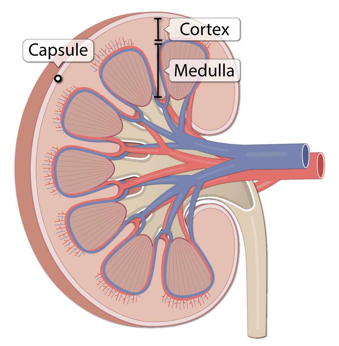
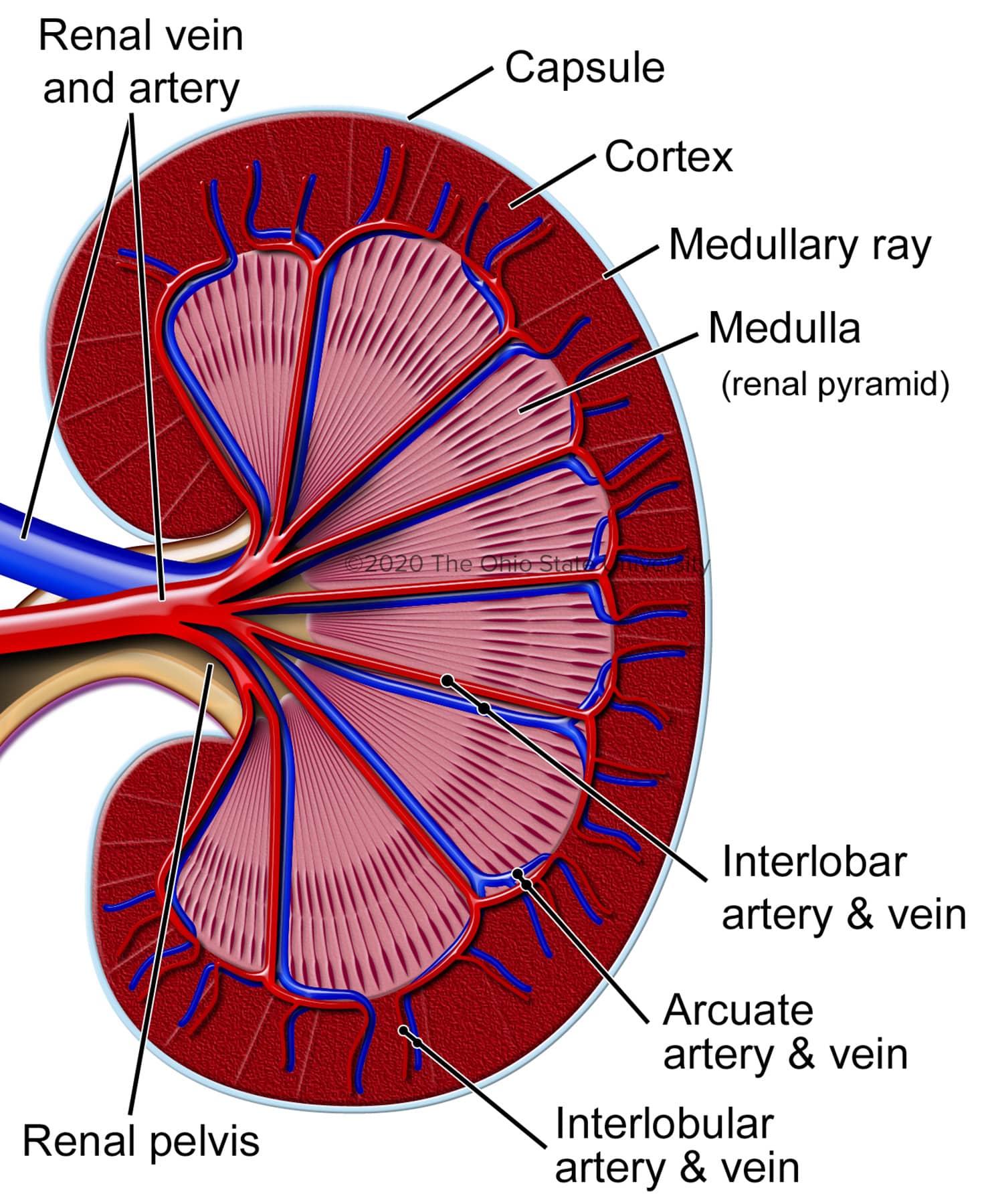
renal medulla: The inner-most region of the kidney, arranged into pyramid-like structures, that consists of the bulk of nephron structure. renal cortex: The outer region of the kidney, between the renal capsule and the renal medulla, that consists of a space that contains blood vessels that connect to the nephrons.
Renal Medullary Vasoconstriction In Normotensive Rats
Fig. 4.Chronic influence of renal medullary interstitial infusion of the nitric oxide synthase inhibitor l-NAME on renal medullary blood flow , daily sodium balance , and mean arterial blood pressure in conscious Sprague-Dawley rats. Vertical dashed lines indicate the l-NAME infusion period. * Significant difference from control .
Recommended Reading: Does Carbonation Cause Kidney Stones
You May Like: Can Kidney Infection Cause Swollen Lymph Nodes
What Are The Three Main Regions Of The Kidney
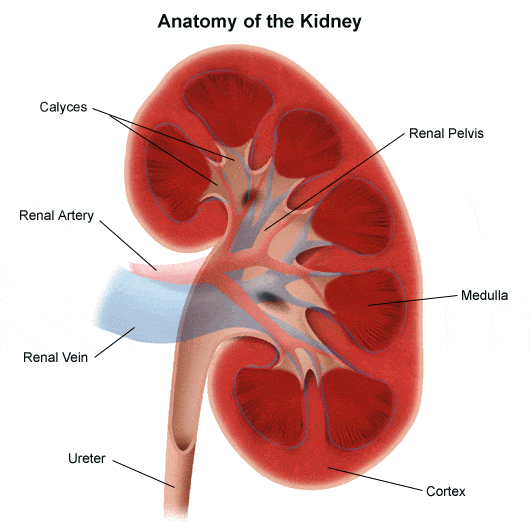
The kidney is made up of three different regions internally: the outer cortex the middle medulla and the inner-most renal pelvis.The kidney is made up of three different regions internally: the outer cortex the middle medulla and the inner-most renal pelvis
the funnel-like dilated part of the ureter in the kidney
How Do You Improve Kidney Function
Here are some tips to help keep your kidneys healthy. Keep active and fit. Control your blood sugar. Monitor blood pressure. Monitor weight and eat a healthy diet. Drink plenty of fluids. Dont smoke. Be aware of the amount of OTC pills you take. Have your kidney function tested if youre at high risk.
You May Like: Does Saw Palmetto Damage Kidneys
How The Kidneys Work
Blood is filtered at high pressure to remove glucose, water, salts and urea.
All the glucose, and some water and salts, are reabsorbed back into the blood. Note that urea is not reabsorbed.
Dr Alice Roberts dissects a pigs kidney and explains the structure and function of the kidney and urinary system
Renal Nerve Anatomy/autonomic Innervation
The kidney receives autonomic supply via both the sympathetic and parasympathetic portions of the nervous system. The preganglionic sympathetic nervous innervation to the kidneys arises from the spinal cord at the level of T8-L1. They synapse onto the celiac and aorticorenal ganglia and follow the plexus of nerves that run with the arteries. Activation of the sympathetic system causes vasoconstriction of the renal vessels. Parasympathetic innervation arises from the 10th cranial nerve , the vagus nerve, and causes vasodilation when stimulated.
You May Like: Is Mio Bad For Your Kidneys
Don’t Miss: Where Are My Dog’s Kidneys Located
Where Is The Renal Cortex
The renal cortex is part of your kidney, which itself is part of the urinary tract. Kidneys are located just below your ribcage and behind your belly. Typically, one kidney sits on either side of your spine. The kidneys are located between your intestines and your diaphragm. Each kidney has a tube-like structure called the ureter which connects the kidney to your bladder.
The renal cortex is brownish-red in color. Its the outside part of the kidney. It covers the renal medulla, the inside part of the kidney. The medulla contains little triangular pieces called the renal pyramids. The renal cortex covers the renal pyramid like a cap.
What Is Renal Cortex

Renal Cortex refers to the part of the kidney that contains the glomeruli and the proximal and distal convoluted tubules. It is covered by the renal fascia and the renal capsule. Since renal cortex contains structures of the nephrons, it is considered as a granular tissue. This smooth, continuous layer of the kidney filters blood. This filtration is called ultra-filtration or high pressure-filtration. Renal artery carries high-pressure blood. Glomeruli are the tiny, ball-shaped arteries, which are encircled by a Bowmans capsule. The fluid in the glomeruli blood leaks into the Bowmans capsule but, red blood cells, white blood cells, platelets, and fibrinogens stay inside the blood capillaries. Blood plasma, glucose, salt, and urea are leaked into the nephrons. Glomeruli leak 160 liters of blood in every 24 hours. Most of the fluid is reabsorbed into the blood in the renal medulla.
Figure 1: Kidney
Proximal and distal convoluted tubules are also found in the renal cortex. The re-filtration of glucose occurs in the proximal convoluted tubules while the distal convoluted tubules re-filter salts. Renal cortex also provides space for arterioles and venules as well as glomerular capillaries. Renal cortex produces hormones called erythropoietin, which facilitates the synthesis of new red blood cells.
Recommended Reading: How Will You Know When A Kidney Stone Passes
Diagnosis Of Medullary Sponge Kidney
Since medullary sponge kidney may not cause symptoms, the condition is often diagnosed during medical investigations for other problems. The presence of kidney cysts and kidney stones may suggest medullary sponge kidney. However, conditions other than medullary sponge kidney can cause kidney stones , so these must be ruled out before medullary sponge kidney is diagnosed.
Tests used to diagnose medullary sponge kidney may include:
- renal ultrasound scan of the kidneys. This is normal in medullary sponge kidney unless stones have formed
- computed tomography scan to detect the presence of cysts, if other tests are inconclusive or if more information is needed.
Kidney stones may be seen in the bladder, ureters or kidneys. In severe cases, imaging may reveal multiple large cysts and clusters of broad kidney stones.
Orexin Regulation Of Sympathetic Centers And Responses
The RVLM and rostral ventromedial medulla appear to be two critical efferent targets for ORX sympathoexcitatory effects . The RVLM plays a critical role in cardiovascular reflexes associated with MAP and in increasing MAP in response to hypertensive stress . Consistent with a role for the RVLM in PeF/DMH-mediated cardiovascular responses is the finding that pressor responses, elicited from disinhibition of the PeF/DMH, can be severely attenuated by microinjecting the GABAA receptor agonist muscimol into the RVLM . Injecting ORX-A and ORX-B into the RVLM elicits not only pressor responses but also tachycardia in many cases . ORX depolarizes many RVLM neurons, predominantly through the ORX2 receptor but also through the ORX1 receptor. Huang and colleagues also show that intracisternal ORX2 receptor antagonists are much more effective than ORX1 receptor antagonists on blocking ORX-A-induced depolarizations and intracisternal ORX-A-induced pressor and tachycardia responses . We have noted significant attenuation of anxiogenic drug , anxiogenic stimuli , and interoceptive stress -induced cardioexcitation with systemic administration of an ORX1 receptor antagonist , but have not tested ORX2 receptor antagonists. However, the above noted studies in this section suggests that ORX2 receptor antagonists may be even more effective than ORX1 receptor antagonists in blocking cardiovascular responses following stress.
Wanda M. Haschek, … Matthew A. Wallig, in, 2010
Don’t Miss: Do You Have Two Kidneys
Hormones Of The Adrenal Glands
The role of the adrenal glands in your body is to release certain hormones directly into the bloodstream. Many of these hormones have to do with how the body responds to stress, and some are vital to existence. Both parts of the adrenal glands the adrenal cortex and the adrenal medulla perform distinct and separate functions.
Each zone of the adrenal cortex secretes a specific hormone. The key hormones produced by the adrenal cortex include:
Advantages Of The Model
One of the advantages of a computational model such as that presented in this study is that it is based on a realistic 3D geometry of the rat renal medulla. The 3D geometry can capture the complexity of the renal geometry that simplified 1D or 2D models cannot. For example, the porosity of DVR and AVR depends on the rate of change in medullary cross-sectional area and the rate of DVR turning. The change in medullary cross-sectional area along the CM axis is very nonuniform and difficult to measure or estimate, so it is very difficult to properly represent the actual change in the cross-sectional area, the rate of vasa recta turning, and so the rate of change of porosity in a simplified 1D or 2D model. The use of realistic 3D geometry helps to overcome this significant issue.
Another advantage of our model is that one can easily modify parameters that cannot be manipulated in experiments. For example, the effect of shunting on renal oxygenation at different parts of the renal medulla cannot be determined experimentally, because shunting cannot be turned off in experiments. One can perform any number of similar virtual experiments using the model to investigate the changes in renal oxygenation under different hemodynamic and/or metabolic conditions. Such virtual experiments could provide valuable insight into where potential hypoxic injury may occur in the kidney.
Don’t Miss: How Long Should It Take To Pass A Kidney Stone
Where Is The Medullary Pyramid In The Kidney
Renal pyramids are kidney tissues that are shaped like cones. Another term for renal pyramids is malpighian pyramids. Between seven and eighteen pyramids exist in the innermost part of the kidney, which is called the renal medulla, in humans, there are usually only seven of the pyramids.
What is the main function of the medullary pyramid?
The medullary pyramids are two white matter formations in the medulla oblongata of the brainstem that carry motor fibres from the corticospinal and corticobulbar tracts, which are commonly understood as the pyramidal tracts.
Which of the following is found in the medullary pyramid of kidneys?
These medullary pyramids contain all the contents of the medulla i.e., transporting tubules like PCT, DCT, and the collecting ducts. It also gets its blood supply from interlobular arteries which divide into the smaller capillaries called peritubular capillaries to form a meshwork of a rich network of blood supply.
Where are the renal pyramids located within the kidney quizlet?
Where are the renal pyramids located? Within the renal medulla, deep to the cortex, projecting to the center.
What makes up the medullary pyramid?
fiber bundles in the medulla that appear triangular in cross-section and contain motor fibers, the majority of which are part of the corticospinal tract. These motor fibers decussate at the base of the pyramids in the pyramidal decussation.
Animal Experiment And Sample Collection
All animal experimental protocols were according to the guide for the care and use of laboratory animals released by the National Research Council of the National Academies, and all experimental protocol was approved by the Animal Ethics Committee of China Pharmaceutical University . Male Sprague-Dawley rats, six to seven weeks old , were allowed to acclimatize for a week. All rats were fed with a standard commercial diet while housed in a light- and temperature-controlled room .
At the first day after acclimatization, rats were intravenously administered with a single dose of cisplatin, and the dosages were 2.5mg/kg , 5.0mg/kg and 10.0mg/kg . The dosages were made according to converted human dosage , lethal dose of 50% in rats , our pre-experiment results and existing literature. Rats in control group was intravenously administered with an equivalent volume of normal saline.
At the seventh day after dosing, rats were sacrificed after blood collection. The sampling time was made combining results of our pre-experiment and existing literature,,,,. The left kidneys were removed, weighted and dissected on an ice plate to separate the cortical and medullar part. All the samples were kept at 80°C until metabolomics analysis. The right kidneys were removed, weighted and cut into two portions. One portion was fixed in 10% neutral-buffered formalin for hematoxylin and eosin staining and another in 4% neutral-buffered paraformaldehyde for TUNEL assay.
Also Check: How Many Calories In A Steak And Kidney Pie
Medullary Changes And Corticomedullary Differentiation
The medullary pyramids may be more prominent in many cases of parenchymal disease as the increased cortical reflectivity increases contrast with the echo-poor medullary tissue. In other cases there may be a decrease in the degree of corticomedullary differentiation so that the pyramids are poorly defined or even indistinguishable as separate structures . However, as with cortical reflectivity, there is no correlation with the aetiology of the renal disease.3 In acute conditions involving primarily the medulla, such as acute tubular necrosis, the pyramids can be enlarged due to oedema. Increased medullary reflectivity can be detected in nephrocalcinosis of any aetiology and also in some other conditions such as gout.
-
Renal US shows echogenicity of medullary pyramids in neonate with normal renal function
-
Usually involves tips & central portions of pyramids
-
Bases of pyramids typically remain hypoechoic
-
Diffuse involvement of entire medullary pyramid uncommon
-
May appear as layering gradient of echogenicity, brightest at tips of pyramids
-
Posterior acoustic shadowing typically absent
-
Segmental involvement > diffuse
-
Normal renal cortical echogenicity & size
Nyree Griffin MD FRCR, Lee Alexander Grant BA FRCR, in, 2013
Read Also: Is Almond Milk Bad For Your Kidneys
Nephrotoxicity Induced By Cisplatin

Body weight, kidney coefficient, blood urine nitrogen and serum creatinine of rats in different groups have been reported in our previous study. The results demonstrated that low-dose cisplatin induced slight kidney damage, and medium- or high-dose cisplatin induced significant renal function decline. It should be noted that the initial numbers of rats were 13, 13, 16 and 26 for group C, L, M and H, respectively. Eventually, 2, 3 and 10 rats in group L, M and H died, and the final numbers of animals were 13, 11, 13 and 16 for group C, L, M and H, respectively.
Not only the macroscopic indicators but also microscopic morphology were significantly changed after cisplatin administration. showed the representative pathological examination results of cortex and medulla in different groups. No abnormal changes were observed in control group , and only slight tubular expansion could be observed in medulla in low-dose cisplatin group . But remarkable abnormal histological changes can be observed in both cortical and medullar part in medium-dose cisplatin group and high-dose cisplatin group . The damages manifested tubular necrosis, tubular expansion, renal epithelial casts and interstitial infiltration of inflammatory cells.
Figure 1: Nephrotoxicity was induced by cisplatin.
You May Like: Celery Juice Kidney
Also Check: Does Anemia Cause Kidney Problems