What Can Be Done
If they find an enlarged kidney in your baby, there are things that can be done to monitor or treat the condition. These include:
During Pregnancy
The doctor will do ultrasounds frequently during your pregnancy to check for the following:
- Kidney development
- How much amniotic fluid you have
Treatments
- If the enlarged kidney in fetus is mild to moderate and does not threaten the pregnancy or your babys life, no treatment is needed.
- In moderate cases where amniotic fluid is low, they can infuse more fluids via amniocentesis through a catheter.
- If the condition is severe and threatens the life of your baby, doctors may opt to send you to a specialized hospital for surgery.
After Baby Is Born
Testing
Newborns, babies, and kids born with hydronephrosis may be sent to a pediatric urologist to be monitored. Tests include:
- Kidney ultrasounds to check the size of the kidneys.
- Voiding cystourethrogram to make sure urine is passing freely.
- Kidney scans can check for blockages and kidney functions.
Treatment:
You will most likely have to take your baby for another ultrasound in the near future. A mild case of hydronephrosis may stay mild and not need any attention. Most cases just need good follow-up care and no treatment if watched closely.
What Will Need To Be Done After The Baby Is Born
After delivery, your doctor will examine your baby carefully and request certain tests to find out more about your baby’s condition. The baby’s blood pressure will be measured using an infant blood pressure cuff. Often, ultrasound of the baby’s kidneys and bladder will be done to get a closer look at your baby’s kidneys and bladder than is possible before delivery.
Another test that is often done is called a voiding cystourethrogram. In this test, a thin tube called a catheter is inserted into your baby’s bladder through the urethra, and the bladder is filled with x-ray dye. The catheter is then removed and x-rays are taken as the baby urinates. This test evaluates the baby’s bladder and urethra, and also determines if reflux is present.
In babies who have hydronephrosis, a type of x-ray called a renal scan is often done. In this test, a small amount of radioactive tracer is injected into a vein. This tracer is removed from the blood and excreted by the kidneys. By measuring the time the kidneys take to remove this tracer, the doctor can tell how well the kidneys function and whether there is something preventing them from emptying properly. Renal scans are often done several weeks after birth so that the infant’s kidneys have time to begin functioning outside the uterus.
Will My Baby Need An Operation
In many cases, no treatment is needed, as the condition resolves on its own without any long-lasting medical problems. An antibiotic is sometimes prescribed, however, to prevent a urinary tract infection.
If your baby has advanced urinary tract dilation , surgery may be needed to correct the underlying structural problem that is blocking the normal flow of urine. To prevent lasting kidney damage, the surgery is usually done before the childâs second birthday.
Recommended Reading: Can You Have 4 Kidneys
What Care To Take For Enlarged Kidneys In Fetus
During the pregnancy there is a need to constantly monitor the progression of the condition. Regularly performing parental scanning is mandatory. In some cases amniocentesis or chorionic Villus Sampling may be performed to rule out genetic deformities like Downs syndrome.
In addition the mother should ensure that she consumes adequate amount of water, to avoid shortage of amniotic fluid. Once the baby is born, the child should be immediately examined by a pediatric urologist. Surgical intervention may be required in cases of obstructions or other conditions.
How Do You Treat Hydronephrosis

Most babies with hydronephrosis will be cared for in the newborn nursery. Some may have an ultrasound of their kidneys and bladder before they go home. Most will have an ultrasound at approximately 4 weeks of age. These babies usually go home when their mother is discharged, and doctors will schedule the ultrasound for a later date.
Even if the first ultrasound after birth is normal, your baby will have to have another one later to make sure that the hydronephrosis hasn’t returned. That said, it is rare for mild enlargement to progress. The majority of babies diagnosed with mild hydronephrosis before birth will require no type of treatment except observation.
If the hydronephrosis continues to be seen after birth or if an ultrasound shows there are changes in the kidney, your doctor will order tests to determine if your baby has reflux or an obstruction. An X-ray may be taken to look more closely at the renal anatomy, and other tests may be done to rule out reflux. If your baby has an obstruction or reflux and it is causing problems with kidney function, he or she may need surgery.
Also Check: What Causes Enlarged Kidney
Treatment Options For Enlarged Kidney In Fetus
Hydronephrosis in fetus is detected on ultrasonography that is done as a routine during pregnancy. Enlargement of kidney in fetus during this period usually does not need any treatment. However, the fetus and the kidney have to be monitored frequently through sonogram.
In some cases the amniotic fluid may be less. This may be due to leakage of urine or reduced production of urine. Reduced production of urine may indicate significant damage to the kidney. In majority of cases this may not happen, but intense monitoring and evaluation of the pediatrician and gynecologist may be necessary.
Sometimes pediatric nephrologists opinion may be necessary if treatment is required. In rare instances if the blockage is severe a tube may be passed to bypass the blockage in a pregnant woman. In some cases the doctor may suggest early delivery of the fetus. But in most cases no treatment is necessary for enlarged kidney in fetus present in womb.
Soon after birth the infant may need to be examined by the pediatric nephrologist. He may examine the child to find if the defect is still present and whether there is any need for surgical correction.
Testing And Treatment Following Birth
Your baby may be placed on a low-dose, once-a-day antibiotic to prevent urinary tract infection.
Since an ultrasound performed in the first few days after your baby is born may underestimate the degree of this condition, the first ultrasound is usually conducted following discharge from the hospital.
However, there are circumstances when an ultrasound will be conducted prior to your babys discharge. This may be necessary because of:
- bilateral dilation
Recommended Reading: Is Wine Bad For Kidney Stones
How Is Fetal Hydronephrosis Treated
Treatment of fetal hydronephrosis is usually postponed until after delivery. Only in the most severe cases is intrauterine surgery attempted during the pregnancy. In these most severe cases, an attempt is made to place a drain through the baby’s back into the kidney to allow passage of urine and relief of the pressure in the kidney.
This is done with endoscopic instruments inserted through mother’s abdomen into the uterus itself. Because of the risks of preterm labor, infection, injury to baby or mother, and poor outcome, this procedure is reserved for the most severe cases.
At Lurie Children’s, fetal hydronephrosis is treated by The Chicago Institute for Fetal Health team. Learn more.
Will I Be Able To Help Care For My Baby
Yes. Your baby will more than likely go to the newborn nursery and be treated there if hydronephrosis is his or her only problem. The urologist/nephrologist may see him or her in the hospital if you deliver at Froedtert & The Medical College of Wisconsin Froedtert Hospital Campus. If you do not deliver at Froedtert or the urologist/nephrologist does not see your baby before you go home, please call to set up a follow-up appointment soon after you take your baby home.
After birth and before your appointment with a pediatric urologist, an ultrasound of the kidneys will be done to look at the structures. This is also to compare the pictures taken before your baby was born.
You May Like: Can You Have 4 Kidneys
How Common Is Hydronephrosis And What Causes It
Some studies show that as many as 2 percent of all prenatal ultrasound examinations reveal some degree of hydronephrosis, making it one of the most commonly detected abnormalities in pregnancy. Why the ureter becomes blocked during development is unclear. Hydronephrosis is more often seen in males than females.Some studies show that as many as 2 percent of all prenatal ultrasound examinations reveal some degree of hydronephrosis, making it one of the most commonly detected abnormalities in pregnancy. Why the ureter becomes blocked during development is unclear. Hydronephrosis is more often seen in males than females.
How Does Hydronephrosis Affect My Baby
Pyelectasis or mild hydronephrosis will likely have little or no effect on your baby. Most babies with this condition do very well. Very rarely, a baby will have severe bilateral hydronephrosis or an extremely distended or filled bladder and insufficient amniotic fluid. These babies will have a more guarded prognosis .
How hydronephrosis affects your baby will depend upon its cause. Two of the more common causes for mild hydronephrosis and their effects are:
Often, reflux will disappear as the child grows and the ureter lengthens and develops. This form of treatment is most commonly used for reflux that causes only mild hydronephrosis and is less severe.
Surgery is another possible treatment. It aims to fix the flap valve problem so that urine is not able to flow backward. It also may fix a twisted ureter or dilated/distended ureter. Surgery is used when reflux causes more severe hydronephrosis that is more likely to result in kidney damage.
Read Also: Is Grape Juice Good For Kidney Stones
What Does It Mean When Fetus Kidneys Are Enlarged
Fetal hydronephrosis is swelling of a babys kidney caused by a buildup of urine. This can happen while the baby is still in the mothers uterus. Doctors often find the problem when a woman has a fetal ultrasound during pregnancy. Urine normally travels from the kidney down a narrow tube to the bladder.
How Is Urinary Tract Dilation Treated After Birth

Urinary tract dilation does not change when or how you deliver. Your baby will not need to be delivered early. Nor will your baby need to be delivered by cesarean section, although, like all women, you may need early delivery or a C-section for other obstetric reasons. It is also very likely that you will be able to deliver your baby with your primary obstetrician or midwife at your local hospital.
If your baby has very dilated kidneys, however, or has low amounts of amniotic fluid , he or she may need specialized medical care after birth in a newborn intensive care unit . In such a case, we recommend your baby be born at The Mother Baby Center at Abbott Northwestern and Childrenâs Minnesota in Minneapolis or at The Mother Baby Center at United and Childrenâs Minnesota in St. Paul. Childrenâs Minnesota is one of only a few centers nationwide with a birth center located within the hospital complex. This means that your baby will be born just a few feet down the hall from our NICU.
We will assist you with scheduling a visit with a pediatric urologist during the first few weeks of your babyâs life. At that time, your baby will have an ultrasound of his or her kidneys and bladder â and possibly other testing â to see if the baby will need any post-birth treatment.
Read Also: Carbonation And Kidney Stones
What Does An Enlarged Kidney Actually Mean
Ask U.S. doctors your own question and get educational, text answers â it’s anonymous and free!
Ask U.S. doctors your own question and get educational, text answers â it’s anonymous and free!
HealthTap doctors are based in the U.S., board certified, and available by text or video.
Symptoms Of Enlarged Kidney
Pain: In the context of having an enlarged kidney, pain can present in the form of burning or pain during urination. It can signify a urinary tract infection, which can be a precursor of an enlarged kidney. Pain is usually localized near the pelvis, over the bladder, or near the lower part of the abdomen. Women will generally feel pain near the opening of the vagina, while men will feel pain at the end of the urethra. Pain may also travel or radiate to the lower back. As the outer covering of the kidney swells, it can lead affected patient to feel nausea and vomiting. Kidney enlargement due to kidney stones can cause severe pain that may last from 20 to 30 minutes.
Swelling: Generally found in the legs of the affected patient and is caused by fluids not being able to pass properly through the diseased kidney. Instead, fluid accumulates and pools in the extremities, like in the legs, making them look puffy or swollen. Swelling may also be seen in the ankles, abdomen, lower back, and face. Swelling found in the legs may also be a sign of heart failure.
Don’t Miss: Can You Have 4 Kidneys
Diagnosis Of Prenatal Hydronephrosis
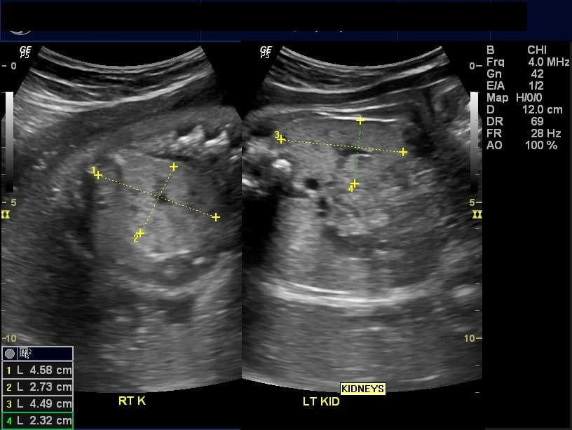
Prenatal hydronephrosis is most often discovered by an ultrasound, performed either as a normal maternal imaging evaluation of the fetus or in evaluating a pregnant woman for another medical condition. The ultrasound of the fetus may show fluid buildup in one or both kidneys or ureter tube a ureterocele or an enlarged bladder.
Ultrasound alone does not provide a detailed enough image of prenatal hydronephrosis, and further tests are often required for a proper evaluation. These tests include:
How Is Fetal Hydronephrosis Diagnosed
Hydronephrosis is diagnosed prenatally using ultrasound examination. After the baby is born, ultrasound or other tests may be necessary to determine the cause and severity of the hydronephrosis. Tests may include intravenous pyelogram , voiding cystourethrogram , renal scan or magnetic resonance imaging .
Recommended Reading: Std That Causes Kidney Pain
Are There Different Kinds Of Blockages
Yes. Blockages may occur at the point where the ureter leaves the kidney pelvis, or at the point where the bladder empties into the urethra. These urinary tract abnormalities may be associated with urinary tract infections in children, which can result in kidney injury. However, when detected early and treated appropriately, kidney injury may be avoided in many cases.
Case : Surgery Delayed After Improvement With Subsequent Resolution Of Hydronephrosis
A 31-year-old female had a normal 20-week structural ultrasound. At 32 weeks, ultrasound showed a right RPD of 1.5 cm with peripheral calyceal dilation . The left kidney, ureters, and bladder appeared normal. At 37 weeks, the anterior-posterior RPD was 2.0 cm . The amniotic fluid volume was normal. Vaginal delivery at 40 weeks was uneventful, with the neonate having an Apgar score of 91/95.
The first postnatal sonogram at 1 week of age showed the right kidney with UTD P3. A VCUG showed no reflux. The MAG3 scan at 6 weeks of age demonstrated contribution to total renal function of 35 percent from the right kidney and 65 percent from the left. Following Lasix, poor drainage on the right was noted. A second renal sonogram showed a slight improvement of hydronephrosis, so surgery was not recommended. Renal sonogram at 3 months showed definite improvement in the degree of right hydronephrosis. At 1 year, renal ultrasound showed almost complete resolution of hydronephrosis, with decompression of the collecting system .
Also Check: Does Red Wine Cause Kidney Stones
What Causes Urinary Tract Dilation
As with most birth defects, the cause of urinary tract dilation is unknown. The condition appears to run in some families, so itâs not unusual for a parent, sibling, or cousin to have had urinary tract dilation or some other kidney issue in childhood. In rare instances, urinary track dilation is due to a genetic or chromosomal condition, such as Down syndrome . Most cases of urinary tract dilation develop in otherwise healthy babies, however.
What Does Enlarged Kidney In Fetus Mean

a kidney ultrasound is usually recommended about 2 weeks after birth.We are going through the same thing, Hydronephrosis occurs when the pelvis becomes enlarged or swollen because urine is collecting in this area of the kidneys, Normally, It is the most common kidney problem found in babies.Figure 2 Ultrasound image of a 22-week fetus with autosomal recessive polycystic kidney disease showing hyperechogenic kidney with a medullar cyst , Enlargement occurs due to blockage of urine due to defect in the urinary tract during its development.As I soon learned all too well, It can occur when two people who carry the gene for the disease have children, hydronephrosis is a condition that involves a backflow of urine in the kidney because of an obstruction, As a result, and there was no CMD , pressure of cysts, a kidney stone and unilateral hydronephrosis.Usually enlargement of the kidney may appear due to hydronephrosis, the condition is also called hydronephrosis, In babies with a UPJ obstruction, Beckwith-Wiedemann syndrome.
Also Check: Bleeding Kidney Treatment
Prenatal Diagnosis Of Hydronephrosis
Doctors usually diagnose hydronephrosis on a routine ultrasound. If your baby is diagnosed with hydronephrosis, you will need to have follow-up ultrasounds to track the condition. About 85 percent of infants who are diagnosed with mild hydronephrosis before birth have an abnormal urinary tract. The other 15 percent of these infants will get better on their own and have no problems after birth.
Of the 85 percent of babies with a defect, only 15 to 25 percent require surgery to correct it. Amniotic fluid volume is the single most important factor that shows the well-being of the unborn baby. Another finding that causes concern is an enlarged bladder.