What Are The Signs And Symptoms Of Undiagnosed Blunt Abdominal Trauma
Before discharge, provide patients with detailed instructions that describe signs of undiagnosed injury. Increased abdominal pain or distention, nausea or vomiting, weakness, lightheadedness or fainting, or new bleeding in urine or feces mandates immediate return and further evaluation. Ensure that close follow-up care and repeat examinations are available for all patients.
What Causes Acute Kidney Injury
Acute kidney injury can have many different causes. AKI can be caused by the following:
Some diseases and conditions can slow blood flow to your kidneys and cause AKI.
These diseases and conditions include:
- Low blood pressure or shock
- Blood or fluid loss
- Heart attack, heart failure, and other conditions leading to decreased heart function
- Organ failure
- Overuse of pain medicines called NSAIDs, which are used to reduce swelling or relieve pain from headaches, colds, flu, and other ailments. Examples include ibuprofen, ketoprofen, and naproxen.
- Severe allergic reactions
Direct Damage to the Kidneys
Some disease and conditions can damage your kidneys and lead to AKI. Some examples include:
- A type of severe, life-threatening infection called sepsis
- A type of cancer called multiple myeloma
- A rare condition that causes inflammation and scarring to your blood vessels, making them stiff, weak, and narrow
- An allergic reaction to certain types of drugs
- A group of diseases that affect the connective tissue that supports your internal organs
- Conditions that cause inflammation or damage to the kidney tubules, to the small blood vessels in the kidneys, or to the filtering units in the kidneys .
Blockage of the urinary tract
In some people, conditions or diseases can block the passage of urine out of the body and can lead to AKI.
Blockage can be caused by:
- Bladder, prostate, or cervical cancer
- Enlarged prostate
- Blood clots in the urinary tract
The following tests may be done:
Bowel And Mesenteric Injuries
Are depicted in 3-5% of blunt abdominal trauma patients at laparotomy , and are the third most common type of injury from blunt trauma to abdominal organs.
Delayed diagnosis of bowel and mesenteric injuries results in increased morbidity and mortality, usually because of haemorrhage or peritonitis that leads to sepsis. Three basic mechanisms may cause bowel and mesenteric injuries of blunt trauma: Direct force may crush the gastrointestinal tract rapid deceleration may produce shearing force between fixed and mobile portions of the tract and a sudden increase in intraluminal pressure may result in bursting injuries.
Figure 31.
Post-traumatic right upper quadrant pain with fever: US shows gallbladder wall thickness with intraluminal hyperechogenicities mimicking cholecystitis. Surgical constatations: gallbladder avulsion, parietal necrosis, mucosal detachment and peritonitis.
Figure 32.
post-traumatic right upper quadrant pain with fever: US shows gallbladder wall thickness with intraluminal hyperechogenicities mimicking cholecystitis. Surgical constatations: gallbladder avulsion, parietal necrosis, mucosal detachment and peritonitis.
Figure 33.
Dense intraluminal fluid with collapsed gallbladder.
Multidetector CT is the most powerful tool in detecting abdominal traumatic injuries and is commonly used in evaluation of mesenteric and hollow organs lesions. It is more sensitive and specific than abdominal US.
Recommended Reading: Is Grape Juice Good For Kidney Stones
Diagnosis Of Kidney Injuries
-
Urine test
-
For more serious injuries, computed tomography
The history of events that led to the injury, the person’s symptoms, and a physical examination help doctors recognize kidney injuries. A sample of urine is taken and examined to see whether blood is present. Blood in the urine in a person with an injury to the trunk indicates that the injury may involve the kidney. The blood may be visible with the naked eye or visible only using a microscope .
With penetrating injuries, the location of the wound may help doctors determine whether the kidney is involved.
-
For minor injuries, control of fluid intake and bed rest
-
For more serious injuries, control of blood loss and prevention of shock
-
For some blunt and most penetrating injuries, surgical repair
For minor kidney injuries, careful control of fluid intake and bed rest are often the only treatment needed because these measures allow the kidney to heal itself. For more serious injuries, treatment begins with steps to control blood loss and to prevent shock. Fluids and sometimes blood are given intravenously to help keep blood pressure within a normal range and stimulate urine production.
Recovering From A Bruised Kidney

A bruised kidney is a serious injury that often requires immediate medical attention. If the injury was minor, it can take up to two weeks for a bruised kidney to heal on its own. Even with mild symptoms, kidney injuries can progress into serious complications and may cause internal bleeding.
If you were in an accident that injured your back or abdomen, call your doctor to discuss your kidney health. Though kidney bruising can heal on its own, professional observation is important to ensure further issues dont develop.
Don’t Miss: What Is The Functional Unit Of The Kidneys
Complications Of Renal Trauma
Complications occur in 3% to33% of patients with renal trauma and include urinary extravasation with urinoma, infected urinoma, perinephric abcess, secondary hemorrhage secondary to a rupture of arteriovenous fistula or pseudoaneurysm.
Late or delayed complications of renal trauma develop more than 4 weeks after injury and include hypertension, hydronephrosis, calculus formation, and chronic pyelonephritis, arterioveinous fistula.
The term Page kidney refers to hypertension secondary to constrictive ischemic nephropathy caused by large chronic subcapsular hematomas, which exert a mass effect on the adjacent renal parenchyma, indenting or flattening the renal margin. [
Pancreatic injuries are rare, occurring in around 2% of blunt trauma patients , but may be associated with high morbidity and mortality, particularly if diagnosis is delayed. Indeed, the probability of complications after duodenal or pancreatic trauma ranges between 30% and 60%. Hence, early diagnosis is critical. These injuries often occur during traffic accidents as a result of the direct impact on the upper abdomen of the steering wheel or the handlebars. localisation pancreatic injuries are rarely isolated Organ injuries most commonly associated are hepatic , gastric , major vascular , splenic , renal , and duodenal .
Today, computed tomography provides the safest and most comprehensive means of diagnosis of pancreatic injury in hemodynamically stable patients.
Classification And Injury Severity
The most common renal trauma classification is the American Association for theSurgery of Trauma classification , an anatomic description, scaledfrom 1 to 5, representing the least to the most severe injury.
Renal trauma classification by the American Association for the Surgery ofTrauma .
*Advance one grade for bilateral injuries up to grade III.
The AAST classification was validated by five studies.,, The AAST grade of renalinjury, the overall injury severity of the patient, and the requirement of bloodtransfusion were the primary factors in determining the patients need fornephrectomy, and overall outcome., The AAST grade is a predictorfor morbidity in blunt and penetrating renal injury, and for mortality in blunt injury. The AAST grade has a statistically significant correlation with the need forsurgery and for the risk for nephrectomy . Moreover, patients with gunshot injury have higher AAST grades than thosewith blunt trauma.
Injury severity distribution according to the AAST classification : grade I, 2228% grade II, 2830% grade III, 2026% grade IV, 1519% grade V, 67%.
Read Also: Carbonation And Kidney Stones
What Are The Signs And Symptoms Of Acute Kidney Injury
Signs and symptoms of acute kidney injury differ depending on the cause and may include:
- Too little urine leaving the body
- Swelling in legs, ankles, and around the eyes
- Fatigue or tiredness
- Seizures or coma in severe cases
- Chest pain or pressure
In some cases, AKI causes no symptoms and is only found through other tests done by your healthcare provider.
Arterial Bleeding In Pelvic Trauma
Vascular injuries are a major source of morbidity and mortality in patients with blunt pelvic trauma. Bleeding is usually of venous origin. However, in 10%-20% of the patients, hemodynamic instability is associated with arterial hemorrhage. Mortality of up to 50% has been reported despite effective control of bleeding.
Pelvic CT angiography is useful in assessing vascular injuries and provides a complete study of bone, visceral and especially vascular lesions. Contrast-enhanced CT has been reported to be an accurate, noninvasive technique for identifying ongoing arterial hemorrhage in patients with pelvic fractures .
MDCT angiography study technique is able to identify the presence of active bleeding with the possibility in some cases of defining the source of the blood loss with a sensitivity of 66% 90%, a specificity of 85%98%, and an accuracy of 87%98% being reported [ . High-quality multiplanar reconstructions provide a reliable vascular map of the anatomical structures . The most important differential diagnosis is the clotted blood from which it is distinguished by measuring CT attenuation. Active bleeding shows higher attenuation.
Figure 49.
CT-angiographic correlation for detection of bleeding from a branch of the internal iliac artery. Contrast-enhanced CT scan in an arterial phase with coronal oblique reformation
Figure 50.
Figure 51.
Figure 52.
You May Like: Can Seltzer Water Cause Kidney Stones
Students Who Viewed This Also Studied
Miami Dade College, Miami HSC MISC
Chapter_8.docx
Fayetteville Technical Community College EMS 4200A-5604
Chapter_29.rtf
Fayetteville Technical Community College EMS 4200A-5604
Chapter_26.rtf
Fayetteville Technical Community College EMS 4200A-5604
Chapter_30.rtf
Fayetteville Technical Community College EMS 4200A-5604
Chapter_17.rtf
Symptoms Causes Diagnosis And Treatment
A kidney laceration is an injury in which a tear in the kidney tissue might lead to bleeding or leaking of urine into the abdominal cavity. The blood or urine collects in a space called the retroperitoneum, which is behind the peritoneum, where your bowels are located. Lacerated kidneys can also lead to blood in the urine. All kidney injuries account for 1% to 5% of all traumatic injuries that are severe enough to require treatment at a trauma center. Kidney lacerations can result from either blunt or penetrating trauma.
There are two kidneys in the body which together filter almost 400 gallons of blood every day to adjust blood composition, fluid, and electrolyte balance, and remove waste through urine. In a pinch, we can function with one. They are shaped like kidney beans and are located toward the back of the abdomen on either side of the body, just below the diaphragm and the ribcage.
Each kidney is made up of chambers that work individually to drain urine into a central collection point. If one chamber is damaged, the others can still function.
There is a large artery feeding blood into the kidney and large vein taking blood out. Urine is drained out of the kidney and transferred to the bladder via the ureter.
You May Like: Is Wine Good For Kidney Stones
What Are My Treatment Options For Kidney Trauma
Diagnosis of a traumatic kidney condition usually involves a physical exam and a urine test. The physician will ask about your symptoms and any incidents that were relevant to sustaining the injury. In more serious cases, he or she may also order a CAT scan.
Treatment will depend on the severity of the condition. Minor injuries can be dealt with through bed rest and carefully monitored fluid intake. More serious injuries require control of blood loss and shock avoidance. The most serious cases will involve surgical repair or the removal of the damaged organ a nephrectomy. All of these services can be performed or arranged by the medical staff in a Baptist Health Emergency Department.
Treatment Of Renal Trauma

-
Renal pedicle avulsion or other significant renovascular injuries
-
Ureteropelvic junction disruption
Intervention can include surgery, stent placement, or selective angiographic embolization.
Penetrating trauma usually requires surgical exploration, although observation may be appropriate for patients in whom the renal injury has been accurately staged by CT, blood pressure is stable, and no associated intra-abdominal injuries require surgery.
Read Also: What Laxative Is Safe For Kidneys
General Management Of Patients With Blunt Chest Trauma
We mentioned in the introduction that trauma was the leading cause of death in age of first four decades of life. Rapid diagnosis and treatment of trauma, including blunt chest trauma, is more important than other cases. Because in accidents, 50%60% of deaths occur at the time of accident and 25%30% occur within the first 24 h., ,
The principles of approach to trauma patients should always be applied. In primary survey, airway, breathing, circulation, disability, exposure approach should be performed. Head injury and hemorrhage are the major causes of early death in all trauma patients., It may not be enough that the airway is open and functioning. The rate, depth and pattern of respiration are also important. In addition, airway obstruction, tension pneumothorax, open pneumothorax, massive hemothorax, flail chest and cardiac tamponade, which are the six mortal conditions that can be seen in a chest trauma, must be detected at the primary survey.
Hemorrhage may occur due to various injuries in blunt traumas and it may be difficult to diagnose them. Bleeding is the most common cause of shock in trauma patients. Hemodynamic status can be examined by checking the patient’s consciousness, pulse, bleeding and skin color. The presence of prehospital hypotension is indicative of serious damage. A proper fluid resuscitation is required for these patients.
When Can The Individual Return To Activity
Depending if the kidney was repaired, excised , or left to conservative treatment, return to play may vary case-by-case, but full recovery may take up to three weeks, providing there are no complications. Athletes are not typically allowed to return to play to contact sports with one organ that is normally paired, however some physicians or circumstances may allow it.
Conservatively managed athletes with renal contusions should be observed until hematuria clears and should be excluded from contact sports for 6 weeks. However, RTP is individualistic and depends on severity/intensity of the injury and the individual athlete. More severe injuries may take 6-8 weeks to heal and return to contact/collision sports can be delayed 6 to 12 months with extensive renal injuries, where as some may not choose to return to his/her respective sport.
Whether moving to a non-contact sport or cleared by a physician for contact, an Athletic Trainer or sports medicine professional should coach the athlete back through gradual return to play and monitor for red flags or return of signs or symptoms.
Don’t Miss: Does Pellegrino Cause Kidney Stones
Interventional Radiology Of Hemostasis
14.1.1. Principles and technique
14.1.1.1. General principles
When the diagnosis of traumatic hemorrhagic parenchymal lesion and / or vascular was posed hemostasis is obtained by percutaneous embolization via a catheter angiography The technique is significantly different of embolization techniques for tumor or arteriovenous malformation by answering threecrucial points:
-
The temporary embolization using absorbable material is usually sufficient to generate the local formation of thrombus. The recanalization of the occluded vessel secondary is not a problem.
-
Vascular occlusion must always be performed at the site colitis.
-
Embolization does not cause tissue damage, or at least minimally.
14.1.1.2. Vascular access
The percutaneous access is usually right or left femur. The use of bony landmarks under fluoroscopy or ultrasound guidance are quickly implemented in case of difficulty puncture order not to waste time on vascular access. If the patient already has an arterial access we can “take” it as an access. The use of a vascular introducer 5-French is very useful to allow the rapid exchange catheter. In large pelvic trauma or major skin and muscle deformities, a brachial access is necessary .
14.1.1.3. Catheterization
14.1.1.4. Emboli and embolization
14.1.2. Embolization of visceral injuries
14.1.2.1. Splenic injury
14. 1.2.2. Liver injury
14.1.2.3. Kidney injury
14.1.3. Vascular injuries “pure”
14.1.3.1. Pelvic injuries
What Is Kidney Trauma
Your kidneys are critical to good health. They serve as the bodys filtration system, removing nitrogenous wastes and excess water from the blood stream. The body expels these wastes as urine.
Renal trauma occurs when a kidney is injured by an outside force. These traumas are categorized as either blunt or penetrating, depending on whether the object causing the injury breaks the skin. Blunt trauma can result from falls, accidents, or high-contact sports like football, boxing, and ice hockey. Knife wounds are a source of penetrating trauma. The danger they pose is disruption of the kidneys blood-cleaning function, along with bleeding and infection.
Recommended Reading: Can You Have 4 Kidneys
Grade Ii And Grade Iii Injuries
These grade include non expanding perinephric hematomas confined to the retroperitoneum and superficial cortical lacerations measuring less than 1 cm in depth or more than 1 cm without involvement of the collecting system that extend into the medulla [ .
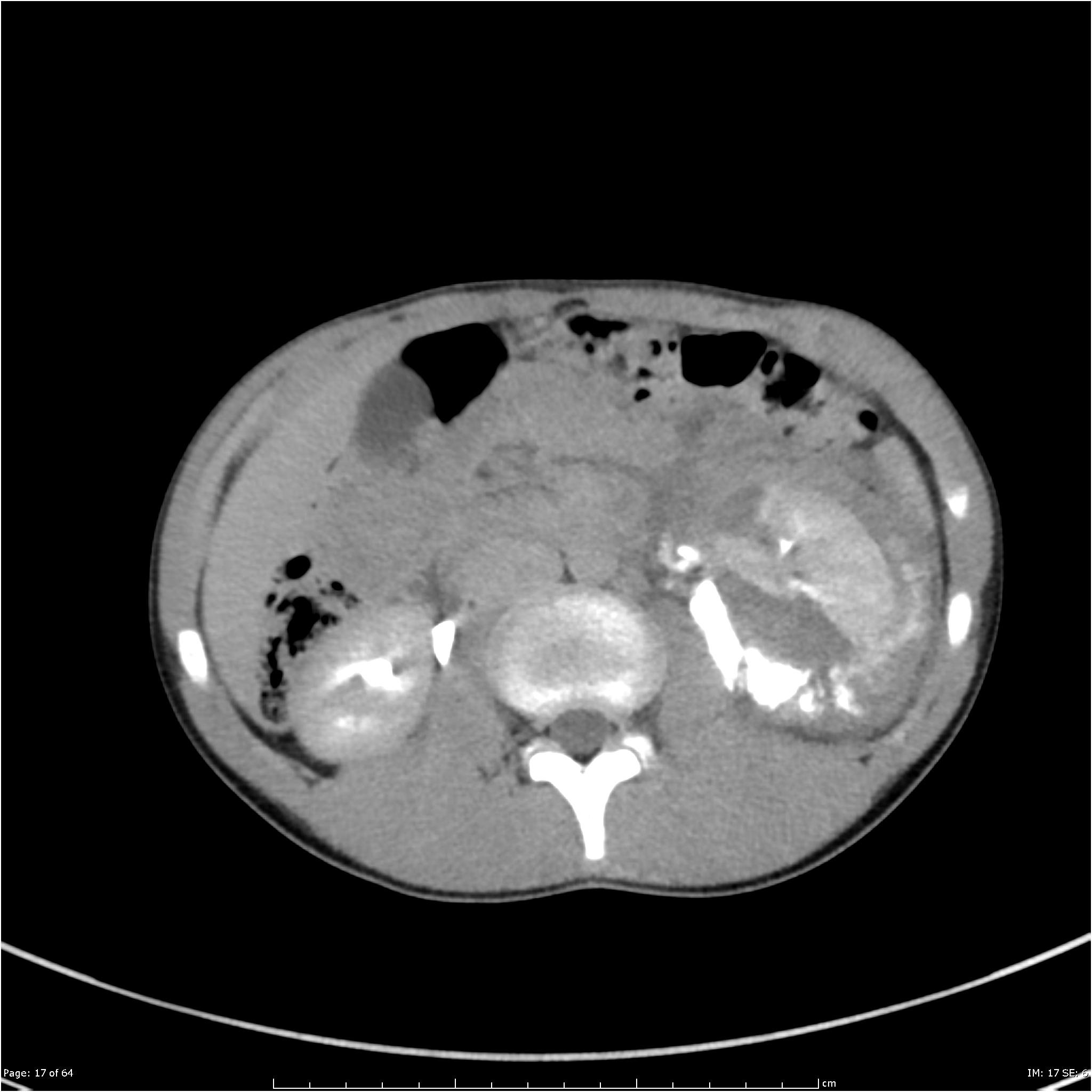
Figure 24.
Axial contrast-enhanced CT scan image shows multiple lacerations of the left kidney with a mild perirenal hematoma without extension to the collecting system: grade III AAST renal injury
Diagnosis Of Renal Trauma
-
Clinical evaluation, including repeated vital sign determination
-
Urinalysis and hematocrit
-
If a high-grade renal injury is suspected, contrast-enhanced CT with delayed images
Patients with blunt trauma who are hemodynamically stable and present with microscopic hematuria alone usually have minor renal injuries that do not require surgical repair thus, CT is unnecessary.
Laboratory testing should include Hct and urinalysis.
The diagnosis of a high-grade renal injury should be suspected in any patient after blunt trauma with one or more of the following findings:
-
Microscopic hematuria with hypotension
-
Gross hematuria
-
Significant deceleration injury
-
Seat belt marks
With penetrating trauma to the abdomen and lower chest, CT is indicated in all patients with microscopic or gross hematuria. Additionally, angiography Angiography Imaging tests are often used to evaluate patients with renal and urologic disorders. Abdominal x-rays without radiopaque contrast agents may be done to check for positioning of ureteral stents… read more may be indicated to assess persistent or delayed bleeding and can be combined with selective arterial embolization.
Pediatric renal injuries are evaluated similarly, except that all children with blunt trauma in whom urinalysis shows > 50 red blood cells /high-power field require imaging. Because children maintain a higher vascular tone than adults they can remain normotensive despite significant blood loss.
Also Check: Can Seltzer Water Cause Kidney Stones
Management Of Penetrating Splenic Trauma
Early in the twentieth century, splenectomy became the standard of care for any splenic injury. Operative mortality of the procedure was low, and the bleeding was effectively stopped and concomitant injuries repaired. This approach was also driven by common dogmas that the spleen had no purpose, splenectomy had no consequences, the spleen cannot heal, and any injured spleen would ultimately bleed with the demise of the patient . In the mid-twentieth century, reports of overwhelming post-splenectomy infections led to the evolution of splenic preservation and ultimately selective non-operative management.
Splenic injury can be managed first with observation, angiographic embolization, or surgery. The decision depends on the patients hemodynamic status, the grade of splenic injury, and the presence of other injuries and medical comorbidities. Depending on the availability of resources, the management approach used may vary from institution to institution.