Comparison Between Volume Regulation And Osmoregulation
Osmoregulation is under the control of a single hormonal system, ADH, whereas volume regulation is under the control of a set of redundant and overlapping control mechanisms. Lack or excess of ADH results in defined and rather dramatic clinical syndromes of excess water loss or water retention. In contrast, a defect in a single volume regulatory mechanism generally results in more subtle abnormalities because of the redundant regulatory capacity from the other mechanisms. Therefore, excess aldosterone results in a mild volume retention followed by escape and return to normal Na+ excretion, due to the action of the other mechanisms. Similarly, excess ANP produces only a modest decrement in volume, with no persistent abnormality in Na+ excretion. Severe salt-retaining states, such as liver cirrhosis or congestive heart failure, are characterized by activation of all the volume regulatory mechanisms.
Laurie J. Vitt, Janalee P. Caldwell, in, 2013
What Function Does The Nephron Result In Osmoregulation
osmoregulation
. Herein, what function does nephron performs in the process of Osmoregulation?
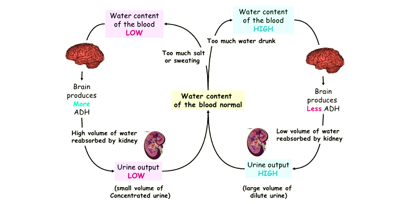
Kidneys play a very large role in human osmoregulation by regulating the amount of water reabsorbed from glomerular filtrate in kidney tubules, which is controlled by hormones such as antidiuretic hormone , aldosterone, and angiotensin II.
Likewise, what are the 4 main functions of a nephron? The nephron uses four mechanisms to convert blood into urine: filtration, reabsorption, secretion, and excretion of numerous substances.
Subsequently, question is, how do nephrons maintain homeostasis?
The kidneys remove waste products from metabolism such as urea, uric acid, and creatinine by producing and secreting urine. Urine may also contain sulfate and phenol waste and excess sodium, potassium, and chloride ions. The kidneys help maintain homeostasis by regulating the concentration and volume of body fluids.
What happens during Osmoregulation?
Osmoregulation is the process of maintaining salt and water balance across membranes within the body. Excess water, electrolytes, and wastes are transported to the kidneys and excreted, helping to maintain osmotic balance. Insufficient fluid intake results in fluid conservation by the kidneys.
If The Relationship Between Reduced Urinary Concentrating Capacity And Hypertension In Adpkd Is Causal What Could Be The Underlying Mechanism
When renal water loss is uncompensated by fluid intake, the ensuing plasma hyperosmolality would stimulate AVP secretion and vasomotor sympathetic outflow. Though an increase in blood pressure may follow, negative feedback mechanisms would maintain normotension. However, in the long term, it is possible that recurrent episodes of hypohydration sensitises blood pressure-regulating neuronal circuits, predisposing individuals to a greater risk of developing hypertension with the presentation of additional temporally separated insults later in life . Conceivably, such an effect could be exacerbated by high dietary sodium. Though this hypothetical mechanism remains to be tested, work done in the Lewis PKD rat has indeed shown a marked chronic upregulation of brain regions that detect and respond to plasma hyperosmolality .
One obvious approach to test whether chronic hypohydration produced by the urinary concentrating defect facilitates the development of hypertension in PKD would be to determine whether increased water intake abrogates the onset or severity of hypertension in patients or animal models. Though the effects of increased water intake on the progression of PKD have been examined, unfortunately hypertension was not included as an outcome measurement for the only clinical trial ; and the single animal study to measure blood pressure was performed on a normotensive rat strain . Hence, these studies do not offer an evaluation of this hypothesis.
Don’t Miss: What Is The Actual Size Of Kidney
The Kidneys And Osmoregulatory Organs
- Explain how the kidneys serve as the main osmoregulatory organs in mammalian systems
- Describe the structure of the kidneys and the functions of the parts of the kidney
- Describe how the nephron is the functional unit of the kidney and explain how it actively filters blood and generates urine
- Detail the three steps in the formation of urine: glomerular filtration, tubular reabsorption, and tubular secretion
Although the kidneys are the major osmoregulatory organ, the skin and lungs also play a role in the process. Water and electrolytes are lost through sweat glands in the skin, which helps moisturize and cool the skin surface, while the lungs expel a small amount of water in the form of mucous secretions and via evaporation of water vapor.
Plasma Membrane Ca2+ Atpase And Epithelial Calcium Channel

In the mammalian distal convoluted tubule, plasma membrane Ca2+-ATPase may mediate Ca2+ transport, thus playing a role in regulating intracellular Ca2+ concentrations . The mammalian PMCA consists of four genes, with PMCA1 and PMCA4 being ubiquitously expressed, while PMCA2 and PMCA3 is more tissue-specific . The pump consists of mainly four domains; the A-domain is important for phosphorylation processes, the P-domain contains the catalytic core of the pump, the N-domain is an important part of the ATP binding site, and finally the calmodulin-binding domain is where the inhibitory binding site is found, freeing it from autoinhibition . The PMCA pump has not been well studied in fish. In the kidney, PCMA isoform regulation could be a important regulatory mechanism of Ca2+ transport. However, Ca2+ regulation related to dietary Ca2+ and environmental levels points to an involvement of both a Na+/Ca2+ exchanger and the Ca2+ ATPase pump in reabsorption in fishes . Further, a decrease in the Ca2+ ATPase enzyme activity in the kidney of tilapia during FW to SW transfer was assumed to reflect a reduced requirement for Ca2+ reabsorption. Although plasma membrane Ca2+ ATPase activity has been measured in gills, intestine and kidney of teleosts potential differential regulation of PCMA isoforms remains elusive.
Don’t Miss: Is Pineapple Good For Kidney Stones
Kidney Function And Physiology
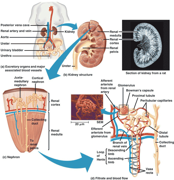
Kidneys filter blood in a three-step process. First, the nephrons filter blood that runs through the capillary network in the glomerulus. Almost all solutes, except for proteins, are filtered out into the glomerulus by a process called glomerular filtration. Second, the filtrate is collected in the renal tubules. Most of the solutes get reabsorbed in the PCT by a process called tubular reabsorption. In the loop of Henle, the filtrate continues to exchange solutes and water with the renal medulla and the peritubular capillary network. Water is also reabsorbed during this step. Then, additional solutes and wastes are secreted into the kidney tubules during tubular secretion, which is, in essence, the opposite process to tubular reabsorption. The collecting ducts collect filtrate coming from the nephrons and fuse in the medullary papillae. From here, the papillae deliver the filtrate, now called urine, into the minor calyces that eventually connect to the ureters through the renal pelvis. This entire process is illustrated in Figure \.
Possible Role For Increased Water Intake In The Treatment Of Pkd
Circulating AVP acts on renal V2 receptors to accelerate cystogenesis in PKD . This was clearly demonstrated by the observation that AVP-deficient PKD rats exhibited a four-fold reduction in cyst volume that was restored upon the administration of a V2 agonist . In the presence of a urinary concentrating impairment, higher plasma AVP would therefore worsen disease progression. Accordingly, clinical trials have investigated the efficacy of a V2 receptor antagonist, Tolvaptan, in ADPKD. Though clinical trials largely recapitulated the positive results shown in animal models, Tolvaptan is not without its side-effects, with safety issues noted relating to increased aquaresis and reduced liver function . Alternative approaches that target the secretion of AVP rather than the V2 receptor are therefore warranted.
Recommended Reading: Is Pineapple Good For Kidney Stones
Osmosensing And Osmosignaling Pathways Toward Canalicular Secretion
Endosomes have been identified as an osmosensing compartment, which is activated in response to hyperosmotic hepatocyte shrinkage . Here, a hyperosmolarityinduced endosomal acidification was shown to trigger ceramide formation within seconds, which in turn activates protein kinase C, which results in an activation of NADPH oxidase isoforms because of an activating phosphorylation of p47phox. As a consequence, hyperosmotic hepatocyte shrinkage produces oxidative stress. It is not yet clear to what extent this oxidative stress response is involved in the hyperosmotic retrieval of canalicular transport systems; however, exogenously added hydroperoxides were shown to induce the retrieval of Mrp2 from the canalicular membrane . In line with the suggestion that oxidative stress may contribute to the cholestatic action of hyperosmotic hepatocyte shrinkage is also the finding that toxic, hydrophobic bile acids, which are known to be cholestatic, also induce oxidative stress via NADPH oxidase activation .
Janet M. Wood, in, 2007
A Possible Link Between Urinary Concentrating Defect And Hypertension In Pkd
There is some initial evidence to suggest that decreased urinary concentrating capacity is linked with the development of hypertension in PKD. Seeman et al. found that the presence of hypertension was 7 times more common in ADPKD children and adolescents who had reduced urinary concentrating ability , and that there was a significant inverse relationship between urine osmolality and ambulatory blood pressure across the cohort. This relationship has also been observed in ADPKD adults. In a retrospective analysis of the TEMPO3/4 study, a large randomised control trial evaluating the efficacy of V2 receptor antagonism in ADPKD, lower urine osmolality at baseline predicted the presence of hypertension . This relationship may partially explain why hypertension associates with indices of cyst abundance , the most likely determinant of the urine concentrating impairment.
You May Like: Does Chocolate Cause Kidney Stones
Summary And Knowledge Gaps In So42 Transport
The reabsorption of SO42 in FW fish is facilitated by an apical SLC13A1 and a basolateral SLC26A1 transporter . In SW fish, SLC26A6A are the most probable candidate for apical transporters involved in SO42 secretion , but SLC26A6B and SLC26A6C may also be involved . The NKA located in the basolateral membrane of proximal tubules provides the driving force for the electrogenic Cl/SO42 exchanger by increasing intracellular negative membrane potential . Finally, a basolateral electroneutral SLC26A1 exchanger for the export of SO42 in exchange for HCO3 probably occurs in both directions for FW and SW fish . There is a need for further studies on the transporters involved in both SO42 reabsorption in FW and secretion in SW, especially characterization of orthologs. Few species have been investigated concerning SO42 transport and several assumptions are still based on the mammalian model. In addition, transport pathways to reabsorb SO42 in FW are still in dispute and have only been verified in the eel.
How Does Osmoregulation Occur In Human Kidney
Kidneyshuman osmoregulationkidneyis
. Keeping this in consideration, where does Osmoregulation occur in the kidney?
Adrenal glands, also called suprarenal glands, sit on top of each kidney. Kidneys regulate the osmotic pressure of a mammal’s blood through extensive filtration and purification in a process known as osmoregulation.
Furthermore, what is human Osmoregulation? Osmoregulation is the control of water levels and mineral ions in the blood. Water levels and mineral ions in the blood are controlled to keep the concentrations the same inside the cells as around them. If the water concentration is too high outside, water enters the cell by osmosis and they may burst.
Likewise, how does Osmoregulation take place in humans?
Osmoregulation is the active regulation of the osmotic pressure of an organism’s body fluids to maintain homeostasis. Kidneys play a very important role in human osmoregulation. They regulate the amount of water in urine waste.
What organs are involved in osmoregulation?
The kidneys are the main osmoregulatory organs in mammalian systems; they function to filter blood and maintain the osmolarity of body fluids at 300 mOsm. They are surrounded by three layers and are made up internally of three distinct regionsthe cortex, medulla, and pelvis.
Recommended Reading: Does Red Wine Cause Kidney Stones
Regulation Of Kidney Function
After answering the question of what do you mean by osmoregulation, now we will learn about the hormones that are responsible for its maintenance. There is hormonal feedback that is responsible for the maintenance of kidney functions. The hypothalamus, the juxtaglomerular apparatus, and the heart are responsible for the regulation of kidney functions. There are certain osmoreceptors that are activated when there are changes in the volume of the blood. When there is excessive loss of fluid from the body then these receptors are activated. The anti-diuretic hormone is released from the neurohypophysis. It is also known as vasopressin. It then facilitates the reabsorption of water from the distal convoluted tubule. When things are normalized, then the secretion of this hormone is stopped. This helps us to understand osmoregulation meaning and the ways by which it is maintained.;
1. Define Osmoregulation?
Ans. The process through which the body maintains osmotic balance throughout membranes is known as osmoregulation. It is an essential biological function.
2. Osmoregulation is Carried Out by Which Organisms?
Ans. Osmoregulation is carried out by all living organisms. Both vertebrates and invertebrates depend on osmoregulation to maintain the overall functionality of their body.;
3. Why is Osmoregulation Necessary in Aquatic Organisms?
Q1. How Does Urea Synthesis Take Place?
Q2. What are Ammonotelism and Ureotelism?
Aquaporins And Their Role In Water Transport In The Kidney Of Euryhaline Fish

The aquaporins, first identified in mammals, constitute several intrinsic proteins that facilitate the passive movement of water molecules across cellular membranes . AQP functions and regulation are complicated, with many aspects yet to be elucidated . Vertebrate aquaporins are heterogeneous, found in diverse tissues, and generally categorized based on their permeability preferences for water, glycerol, and other small solutes . Of the 10 AQP isoforms so far described, 7 are present in the mammalian kidney, located along the nephron and collecting duct, emphasizing their importance for renal water handling .
Several teleost AQPs has been identified and annotated based on their mammalian analogs: AQP-1aa, -1ab, -3a, -3b, -7, -8aa, -8ab, -9a, -10a, 10b, and -12 . Even though several are expressed in the teleost kidney , their roles in renal physiology remain elusive . However, recent investigations have shed some light on AQP1, AQP3, AQP8, AQP10, AQP11 and AQP12 regulation and role in euryhaline fish .
Recommended Reading: Does Red Wine Cause Kidney Stones
What Are Osmoregulation And Excretion
Organisms must keep bodily fluids at a constant temperature and pH while maintaining specific solute concentrations in order to support life functions. Osmoregulation is the process that balances solute and water levels.
Osmosis is the tendency of water to move from solutions with lower ion concentrations, or osmolarities, to those with higher ion concentrations. Osmosis occurs in response to differences in the molecular concentrations of solutions separated by a semipermeable membrane.
Bodily fluids, which are separated by such membranes, contain water, non-electrolytes, and electrolytessolutes that dissolve into ions in water. Both electrolytes and non-electrolytes influence osmotic balance. However, since the more important factor to osmosis is solute number, rather than size, the contribution of electrolytes is more significant.
Unlike water, electrolytes cannot diffuse passively through membranes but rely on facilitated diffusion and active transport. In facilitated diffusion, protein-based channels move solutes across membranes. Conversely, energy is used to move ions against concentration gradients in active transport.
When animals ingest food, material that cannot be used is excreted from the body. Excretory systems in nature involve tradeoffs between conserving energy and water.
Suggested Reading
Please enter your institutional email to check if you have access to this content
If you want more info regarding data storage, please contact .
Thank You
Thank You
Summary Of Aqps And Their Relation To Ion Transport
Engelund and co-authors have contributed extensively to our current understanding of aquaporins in all osmoregulatory organs of euryhaline teleosts, including the possible involvement of other tight junction claudins in paracellular transport of both mono and divalent ions . Nevertheless, the current review highlights the lack of knowledge on the distribution and role of AQPs in water and ion transport in the teleost kidney . The close linkages between the transport of Na+, Cl and water in proximal tubules and reabsorption in distal segments are crucial aspects for future studies. The prevailing hypothesis, at least in euryhaline species, is that AQPs likely serve a fundamental role in transcellular water transport in nephron tubules . However, very few AQPs have been located in the distal tubules and collecting duct of fish where the primary functions are reabsorption of monovalent ions. Presumably, lack of water channels here would be adaptive for life in FW, allowing for minimal water reabsorption and thus facilitate excretion of excess water. In contrast, permeability to water in these segments would be crucial for water absorption in SW, so as to facilitate a low volume of isotonic urine rich in Mg2+, Ca2+ and SO42 . Functional connections between AQPs, claudins, monovalent ion handling , and divalent ion handling are a rich area for future investigation in the kidney of euryhaline fish .
Also Check: Ginger Tea Dissolves Kidney Stones
What Is The Role Of The Kidneys In Osmoregulation
Osmoregulation is the homeostatic control of the water potential of the blood. The kidneys are involved in filtering the blood and deciding which substances to reabsorb and which to excrete as waste. Tiny tubular structures known as tubules carry out this filtration.There are five main parts to the kidney tubules: the Bowman’s capsule, the proximal convoluted tubule, the loop of Henle, the distal convoluted tubule and the collecting duct. The Bowman’s capsule is used for ultrafiltration as the fluid is forced out of the blood and into the tubule. Then, the proximal convoluted tubule is used for the selective reabsorption of all glucose and some salts and water. The loop of Henle then maintains a sodium gradient, before the distal convoluted tubule makes final adjustments to the amount of water and salts that are reabsorbed. The collecting duct is the final piece of the puzzle that allows water to move out by osmosis to decrease the water content of urine.
Paracellular Transport And Tight
Tight junctions proteins may supplement AQP-mediated transporters in water transport where they provide a paracellular route. In the gills a large review has been dedicated to the importance of paracellular permeability with respect to environmental factors . In the kidney of euryhaline teleosts, there is little information on water transport by claudins but CLDN2 have been observed to create water pores in the mouse proximal renal tubules . CLDN2 has not been investigated in the fish kidney. CLDN15a has been identified in salmon, but its permeability properties remains unknown . The authors also discuss the possible involvement of other tight junction claudins in paracellular transport of both mono and divalent ions .
Recommended Reading: What Laxative Is Safe For Kidneys