Safety Of Low Dose Iv Contrast Ct Scanning In Chronic Kidney Disease
| The safety and scientific validity of this study is the responsibility of the study sponsor and investigators. Listing a study does not mean it has been evaluated by the U.S. Federal Government. Read our disclaimer for details. |
| First Posted : June 19, 2015Last Update Posted : November 28, 2017 |
- Study Details
| Phase | ||
|---|---|---|
| Chronic Kidney DiseasePulmonary EmbolismRenal Artery StenosisPulmonary Cancer | Drug: Low Volume iso-osmolar non-ionic radio contrast mediumDrug: Acetylcysteine InhalationDrug: Sodium Bicarbonate SolutionProcedure: 64-MDCT Scanning | Phase 4 |
| Study Type : | |
| Diagnostic | |
| Official Title: | Safety of Low Dose Intravenous Contrast 64 Multi-Detector Computed Tomography Scanning in Patients With Chronic Kidney Disease |
| Study Start Date : |
Impact Of Renal Dysfunction On Diagnostic Imaging Modalities
The frequency of SPECT between the non-decreased eGFR and decreased eGFR groups was 75.9% and 24.1%, respectively , while that of CT was 85.9% and 14.1% and that of CAG was 78.2% and 21.8% . The patient characteristics with each modality in the decreased eGFR group are presented in Table; and are consistent with those in the original J-COMPASS study. In brief, patients who underwent CAG were more likely to be habitual smokers, have peripheral artery or aortic disease, and have high-grade symptoms of angina and dyspnea . After adjusting for confounders, the odds ratio for a higher likelihood to undergo SPECT rather than CT was 1.96 for patients in the decreased eGFR group relative to patients in the non-decreased eGFR group , and the odds ratio for a higher likelihood to undergo CAG rather than CT was 1.56 for patients in the decreased eGFR group relative to those in the non-decreased eGFR group . Renal dysfunction was significantly associated with the choice of initial diagnostic imaging modality.
Table 2 Patients characteristics among SPECT, CT and CAG groups in decreased eGFR group.
Use Of Radiological Tools For Evaluating Kidney Disease
Plain radiographs, or x-rays as they are commonly known, have been in existence for over 100 years, since William K. Roentgen discovered this technology in 1895. Even though there has been an explosion of technology in the imaging modalities, the age-old radiographs still play a role in the diagnosis of chronic kidney disease . The different tools available include plain radiographs , ultrasound, computerized tomography , magnetic resonance imaging and angiography.
The tests available for diagnosing kidney diseases
Plain Radiographs
These films are mainly used during the initial assessment for kidney stones and sometimes to measure the size and shape of the kidney.
Intravenous Urography
IVU is used to measure kidney size and shape and in the evaluation of the pelvis and ureters . The major drawback for this test is the use of contrast dyes, which can have serious side effects including renal failure. Although the risk is less with the newer non-ionic contrasts, the risk still exists.
Angiography
This technique uses contrast dye like the IVU but can provide more information about the blood vessels. The angiogram helps to assess for renal artery stenosiswhen the lining of the main artery supplying blood to the kidney narrows or is blockedlike is used in diagnosing coronary heart disease.
Ultrasonography
Computed tomography
Magnetic resonance imaging
When is the best time to use a particular test?
Acute renal failure
Chronic kidney disease
Renal vein thrombosis
You May Like: Are Laxatives Bad For Your Kidneys
Pet Scanning And Image Processing
The PET component of the PET/CT scanner is composed of lutetium-yttrium oxyorthosilicate-based crystal. Emission scans were acquired at 12 min per bed position. The FOV was from the top-of-head to the bottom of feet in the vast majority of patients. The three-dimensional whole-body acquisition parameters were 128 × 128 matrix and 18 cm FOV with a 50% overlap. Processing used the 3D Row Action Maximum Likelihood Algorithm method. Total scan time per patient was approximately 2045 min.
What Lab Values Indicate A Need For Concern When Contrast Material Is Injected

Historically serum creatinine was the lab value used to assess kidney function. ;A better and more accurate measure is a lab result called estimated glomerular filtration rate . eGFR takes into account the serum creatinine value and also patient age, race and gender which affect kidney function results. ;At UCSF we use this very accurate blood test to assess kidney function and it can be obtained quickly, right before a scan. ;For CT, eGFR > 45 indicates no increased risk of kidney damage from contrast material. ;eGFR > 30, but less than 45 indicates that while it is safe to get contrast material, there is a small risk of causing kidney damage. ;In that situation, we will inject additional fluid into the patients vein before and after the contrast material injection. ;This hydration is effective to prevent any renal damage. ;For MRI, it is safe to give a regular dose of contrast material as long as the patients eGFR is > 30.
Don’t Miss: Is Pineapple Juice Good For Kidney Stones
How Is Kidney Failure Treated
Treatment options vary widely and depend on the cause of kidney failure, but most require a hospital stay. Options are sorted into two groups: treating the cause of renal failure versus replacing the renal function. They include:
- Interventional radiology procedures such as ureteral stenting and nephrostomy: This procedure involves inserting either small stents;into the ureter or a tube connected to an external drainage bag. Both options are used to unblock the ureters in order to allow proper urine flow from the kidneys if this has been identified as the cause for the renal failure.
- Surgical treatment such as a urinary stent or kidney stone;removal.
- Dialysis, including hemodialysis;and peritoneal dialysis: These procedures remove wastes and excess fluid from the blood and therefore replace renal functions. Kidney transplant is the most complete and effective way to replace kidney function but may not be suitable for all patients.
Impact Of Renal Failure On F18
- 1Saint Louis University School of Medicine, Saint Louis, MO, United States
- 2Division of Nuclear Medicine Technology, Saint Louis University Hospital, Saint Louis, MO, United States
- 3Division of Nuclear Medicine, Department of Radiology, Saint Louis University, Saint Louis, MO, United States
Objective: The current guidelines for 2-deoxy-2-fluoro-d-glucose PET/CT scanning do not address potential inaccuracies that may arise due to patients with renal failure. We report a retrospective analysis of standard uptake values in patients with and without renal failure in order to warrant a protocol adjustment.
Methods: Patients were matched based on age, gender, and BMI all of which are potential effectors on observed SUV. Thirty patients were selected with clinically diagnosed renal failure, of which 12 were on dialysis. All 30 patients had age, gender, and BMI control matches. Blood urea nitrogen and creatinine levels were measured within 1 month of the scan to assess renal failure. PET/CT scans for both the renal failure patients and controls were performed 60 min after FDG injection. SUVs were measured by placing circular regions of interest in the right hepatic lobe and left psoas muscle .
Our data suggest that renal failure patients do not require an adjustment in protocol and the standard protocol times should remain.
Read Also: Is Pomegranate Juice Good For Your Kidneys
Imaging Tests To Look For Kidney Cancer
Imaging tests use x-rays, magnetic fields, sound waves, or radioactive substances to create pictures of the inside of your body. Imaging tests are done for a number of reasons, such as:
- To look at suspicious areas that might be cancer
- To learn how far cancer might have spread
- To help determine if treatment is working
- To look for possible signs of cancer coming back after treatment
Unlike most other cancers, doctors can often diagnose kidney cancer with fair certainty based on imaging tests without doing a biopsy . Some patients, however, may need a biopsy.
The Cat Scan Or Ct Scan
A computed axial tomography , or CT scan, produces a series of very detailed cross-sectional views of bones and all types of body tissue, including muscles and blood vessels. A CT scan can show the exact location and size of a tumor, if present, as well as its relationship to surrounding tissue. It is usually the preferred method for detecting cancers and guiding biopsies and related treatments. CT scans can also be used to detect pulmonary embolisms and aortic aneurysms, as well as vascular diseases that can lead to stroke or kidney failure. They can help diagnose injuries to skeletal structures and can detect congenital malformations of the heart and other organs, identify injuries to internal organs, or assess results of organ transplants. For a CT scan, you will typically lie motionless on a CT exam table while a large scanning tube rotates around the table, as the table passes through the tube. The imaging tube scans so quickly less than 30 minutes that even children rarely need to be sedated to remain still during a CT scan. A contrast material may be administered via mouth or IV before your scan.
Also Check: How Much Money Is A Kidney Worth
What Are The Reasons For A Ct Scan Of The Kidney
A CT scan of the kidney may be performed to assess the kidneys fortumors and other lesions, obstructions such askidney stones, abscesses,polycystic kidney disease, and congenital anomalies, particularly when another type ofexamination, such as X-rays or physical examination, is not conclusive.CT scans of the kidney may be used to evaluate the retroperitoneum . CT scansof the kidney may be used to assist in needle placement inkidney biopsies.
After the removal of a kidney, CT scans may be used to locate abnormalmasses in the empty space where the kidney once was. CT scans of thekidneys may be performed afterkidney transplantsto evaluate the size and location of the new kidney in relation to thebladder.
There may be other reasons for your doctor to recommend a CT scan ofthe kidney.
What Is Contrast Induced Nephropathy
CIN is a rare disorder and occurs when kidney problems are caused by the use of certain contrast dyes. In most cases contrast dyes used in tests, such as CT and angiograms, have no reported problems. About 2 percent of people receiving dyes can develop CIN. However, the risk for CIN can increase for people with diabetes, a history of heart and blood diseases, and chronic kidney disease . For example, the risk of CIN in people with advanced CKD below 30 mL/min/1.73m2), increases to 30 to 40 percent. The risk of CIN in people with both CKD and diabetes is 20 to 50 percent.
CIN is associated with a sharp decrease in kidney function over a period of 48-72 hours. The symptoms can be similar to those of kidney disease, which include feeling more tired, poor appetite, swelling in the feet and ankles, puffiness around the eyes, or dry and itchy skin. In many cases, CIN is reversible and people can recover. However, in some cases, CIN can lead to more serious kidney problems and possible heart and blood vessel problems.
Don’t Miss: Can Kidney Stones Cause Constipation Or Diarrhea
Study: Before A Ct Scan Or Angiogram Many People Should Take Inexpensive Drug To Protect Kidneys
Iodine contrast agents that enhance the scans can harm vulnerable kidneys, but N-acetylcysteine taken beforehand can protect at-risk patients
Michigan Medicine – University of Michigan
ANN ARBOR, Mich. As more and more Americans undergo CT scans and other medical imaging scans involving intense X-rays, a new study suggests that many of them should take a pre-scan drug that could protect their kidneys from damage.
The inexpensive drug, called N-acetylcysteine, can prevent serious kidney damage that can be caused by the iodine-containing dyes that doctors use to enhance the quality of such scans.
That dye, called contrast agent, is usually given intravenously before a CT scan, angiogram or other test. But the new study shows that taking an N-acetylcysteine tablet before receiving the contrast agent can protect patients and that it works better than other medicines that have been proposed for the same purpose.
People whose kidneys are already vulnerable, including many older people and those with diabetes or heart failure, are the most at risk from contrast agents, and have the most to gain from taking the drug.
Only N-acetylcysteine clearly prevented contrast-induced nephropathy, the medical name for kidney damage caused by contrast agents. Theophylline, another drug that has been seen as a possible kidney-protecting agent, did not reduce risk significantly. Other drugs had no effect, and one, furosemide, raised kidney risk.
Journal
What Is A Ct Scan Of The Kidney

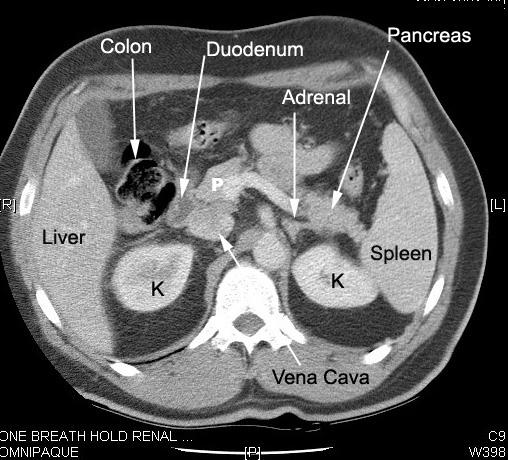
Computed tomography is a noninvasive diagnosticimaging procedure that uses a combination ofX-raysand computer technology to produce horizontal, or axial, images of the body. A CT scan shows detailed images of any part ofthe body, including the bones, muscles, fat, and organs. CT scans are moredetailed than standard X-rays.
In standard X-rays, a beam of energy is aimed at the body part beingstudied. A plate behind the body part captures the variations of the energybeam after it passes through skin, bone, muscle, and other tissue. Whilemuch information can be obtained from a standard X-ray, a lot of detailabout internal organs and other structures is not available.
In computed tomography, the X-ray beam moves in a circle around the body.This allows many different views of the same organ or structure. The X-rayinformation is sent to a computer that interprets the X-ray data anddisplays it in a two-dimensional form on a monitor.
CT scans may be done with or without “contrast.” Contrast refers to asubstance taken by mouth or injected into an intravenous line thatcauses the particular organ or tissue under study to be seen more clearly.Contrast examinations may require you to fast for a certain period of timebefore the procedure. Your doctor will notify you of this prior to theprocedure.
Other related procedures that may be used to diagnose kidney problemsincludeKUB X-rays,kidney biopsy,kidney scan,kidney ultrasound,renal angiogram, andrenal venogram.
Recommended Reading: Is Grape Juice Good For Kidney Stones
Editorial Note On The Review Process
F1000 Faculty Reviews are commissioned from members of the prestigiousF1000 Faculty and are edited as a service to readers. In order to make these reviews as comprehensive and accessible as possible, the referees provide input before publication and only the final, revised version is published. The referees who approved the final version are listed with their names and affiliations but without their reports on earlier versions .
The referees who approved this article are:
-
Ton Rabelink, Department of Nephrology, Leiden University Medical Centre, Leiden, The Netherlands
No competing interests were disclosed.
-
Aiko de Vries, Department of Nephrology, Leiden University Medical Center, Leiden, The Netherlands
No competing interests were disclosed.
-
Ilona Dekkers, Department of Nephrology, Leiden University Medical Center, Leiden, The Netherlands
No competing interests were disclosed.
-
Lei Zhang, Department of Radiology and Imaging Sciences, University of Utah, Salt Lake City, USA
No competing interests were disclosed.
-
Stefan Reuter, Department of Medicine D, University of Münster, Münster, Germany
No competing interests were disclosed.
Can A Ct Scan Detect Polycystic Kidney Disease
CT scan CT is one of the most commonly used medical tests for diagnosing Polycystic Kidney Disease. With CT scan, cysts in kidney can be seen clearly and also this test helps to show kidney shape and enlargement of bilateral kidneys.
And it would be great if there were a safe, effective way to catch the disease.
can be an option. When you might need a CT scan: A scan can be used to diagnose possible appendicitis and kidney.
or polycystic kidney disease is the initial test in most patients. An IVP may be a reasonable first choice in young patients, since it can detect lesions such as medullary sponge kidney that may.
Imaging studies (sonography, computed tomography, magnetic.
The diagnosis of ADPKD usually consists in visualization of multiple cysts in an.
10 H.U. are most likely thick-content cysts (although tumor can’t be ruled out.
A CT scan can make detailed.
congenital anomalies, polycystic kidney disease, buildup of fluid around the kidneys, and the location of abscesses. Your healthcare provider may need to do other.
Chapter XIII.3. Cystic Kidney Disease The infant is taken to the OR for bilateral nephrectomies secondary to autosomal recessive polycystic kidney disease and dialysis catheter placement.
A DMSA renal scan can.
Kidney imaging findings can also vary considerably, depending.
procedures such as CT scans and MRI also can detect cysts.
PKD cysts can slowly replace much of the kidneys, reducing kidney.
Occasionally, a CT scan and MRI.
You May Like: Can Kidney Stones Cause Constipation Or Diarrhea
How Chronic Kidney Disease Is Diagnosed
Chronic kidney disease is primarily diagnosed with blood and urine tests that detect chemical imbalances caused by the progressive loss of kidney function. The tests may be accompanied by imaging tests and biopsies used to pinpoint the exact cause of the dysfunction. Kidney function tests, also known as renal function tests, are important for monitoring the progression of the disease and your response to therapy. They are also vital to staging the disease and can help differentiate CKD from an acute kidney injury .
Subsequent Treatment And Renal Dysfunction
One of the novel findings of the present study is that a decrease in eGFR had an impact on the subsequent treatment strategies. The original J-COMPASS study showed a preference for intervention therapy in patients who underwent CT and CAG compared with those who underwent SPECT, consistently with the findings of the present study. Moreover, renal dysfunction was independently associated with the treatment strategies. One reason for this may be the atherosclerotic burden in patients with renal dysfunction. Another reason may be comorbidities underlying the renal dysfunction, although we performed extensive adjustment for confounding factors.
You May Like: Can Kidney Stones Affect Your Psa Count